
miRNA & the Liver
The liver has the unique capacity to regenerate after surgical resection. The lost functional mass is replaced in a process of compensatory growth in which quiescent hepatocytes reenter the cell cycle. A lot of insights have been gained into the mechanisms of liver regeneration, but the regulation of liver regeneration is not completely understood. Unfortunately, therapeutically induction of liver regeneration still remains difficult.
Recent reports indicated an essential role for small noncoding microRNAs (miRNAs) in the regulation of hepatic development, carcinogenesis, and early regeneration. miRNAs are the most abundant class of small, endogenous noncoding RNAs. miRNAs inhibit protein synthesis by blocking translation via complementary binding of messenger RNA (mRNA) or by suppressing translation and the subsequent degradation of target mRNA. A few studies have already investigated miRNA expression changes during liver regeneration, indicating that miRNA play an important role during liver regeneration.
Recent reports indicated an essential role for small noncoding microRNAs (miRNAs) in the regulation of hepatic development, carcinogenesis, and early regeneration. miRNAs are the most abundant class of small, endogenous noncoding RNAs. miRNAs inhibit protein synthesis by blocking translation via complementary binding of messenger RNA (mRNA) or by suppressing translation and the subsequent degradation of target mRNA. A few studies have already investigated miRNA expression changes during liver regeneration, indicating that miRNA play an important role during liver regeneration.
miRNA & the Liver
We hypothesized that miRNA are critically involved in all phases of liver regeneration. We performed miRNA microarray analyses after 70% partial hepatectomy in rats at different time points (0 h to 5 days) and after sham laparotomy. Putative targets of differentially expressed miRNAs were determined using a bioinformatic approach and two-dimensional proteomic analyses and protein identification were performed on specimens at 0 and 24 h after resection. We observed that miRNA expression patterns changed during liver regeneration and that these changes were most evident during the peak of DNA replication at 24 hours after resection. Expression of 13 miRNAs was significantly reduced 12–48 h after resection (>25% change), whereas three miRNAs were significantly upregulated. Proteomic analysis revealed 65 up-regulated proteins; among them, 23 represented putative targets of the differentially expressed miRNAs.
Our initial study on miRNA expression changes during liver regeneration resulted in a temporal miRNA expression and proteomic dataset of the regenerating rat liver. Our data indicate that multiple miRNAs are involved in regulation of liver regeneration. Thus, we consider the role of miRNA during liver regeneration as being a moderator or conductor rather than a regulator or initiator. We currently perform functional studies to further verify the role of specific miRNA during liver regeneration.
For more detailed information please refer to Am J Physiol Regul Integr Comp Physiol 2011;300:R1363–R1372.
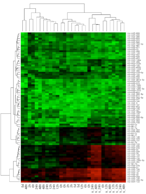
MicroRNA (miRNA) expression during rat liver regeneration after PH.
Representative heat map shows 85 of the 323 investigated miRNA with the highest overall variability. miRNA expression levels at 12, 24, and 48 h after PH are clustered together and are different compared with the other time points and sham controls. Red and green indicate up- and downregulation and the numeric scale bar displays relative intensity values.