News

Spacial computing in the OR
Stacks Image 19721

We tested the Apple Vision Pro in the operating theatre and it cuts an excellent figure: great images even in challenging lighting situations, stable interaction with the device - even though the limited peripheral vision and awareness inherent to video-based devices is a considerable downside in surgery.

We are looking forward to our first software solutions for improved hand-eye coordination in visceral surgery for this device too!
Optimizing environmental enrichment for Sprague Dawley rats: Exemplary insights into the liver proteome
Stacks Image 19730
Considering the expected increase in the elderly population and the growing emphasis on aging-related biomedical research, the demand for aged laboratory animals has surged, challenging established husbandry practices. Our objective was to establish a cost-effective method for environmental enrichment, utilizing the liver as a representative organ to assess metabolic changes in response to differing enrichment levels.
We conducted a six-month study involving 24 male Sprague Dawley rats who were randomly assigned to four environmental enrichment groups. Two groups were housed in standard cages, while the others were placed in modified rabbit cages. Half of the groups received weekly playtime in an enriched rat housing unit. We evaluated hormone levels, playtime behavior, and subjective handling experience. Additionally, liver tissue proteomic analysis was performed.
Initial corticosterone levels and those after 3 and 6 months showed no significant differences. Yet, testosterone levels were lower in the control group by the end of the study (p=0.007). In the liver tissue, we detected 1,871 distinct proteins, with 77% of them being consistent across all groups. In gene ontology analysis, no specific pathways were overexpressed. In semiquantitative analysis, we observed differences in proteins associated in lipid metabolism such as Apolipoprotein A-I and Acyl-CoA 6-desaturase, which were lower in the control group (p= 0.024 and p=0.009). Enriched environments reduced rat distress, large cages eased handling, and conflicts between rats lessened with bi-weekly interactions.

The manuscript "Optimizing environmental enrichment for Sprague Dawley rats: Exemplary insights into the liver proteome" has been accepted for publication in PLOS ONE.
Authors are Nathalie N. Roschke, Karl H. Hillebrandt, Dietrich Polenz, Oliver Klein, Joseph MGV Gassner, Johann Pratschke, Felix Krenzien, Igor M. Sauer, Nathanael Raschzok, and Simon Moosburner.
Proteomic analysis of decellularized mice liver and kidney extracellular matrices
Stacks Image 19695
Based on the collaboration between the Department of General, Visceral, and Transplant Surgery, University Hospital Münster, and Experimental Surgery, Department of Surgery, Charité – Universitätsmedizin Berlin our work on the "Proteomic analysis of decellularized mice liver and kidney extracellular matrices" has been accepted for publication in Journal of Biological Engineering.

In this study, we employed a bottom-up proteomic approach to elucidate the intricate network of proteins in the decellularized extracellular matrices of murine liver and kidney tissues. This approach involved the use of a novel, perfusion-based decellularization protocol to generate acellular whole organ scaffolds. Proteomic analysis of decellularized mice liver and kidney ECM scaffolds revealed tissue-specific differences in matrisome composition, while we found a predominantly stable composition of the core matrisome, consisting of collagens, glycoproteins, and proteoglycans. Liver matrisome analysis revealed unique proteins such as collagen type VI alpha-6, fibrillin-2 or biglycan. In the kidney, specific ECM-regulators such as cathepsin z were detected. The identification of distinct proteomic signatures provides insights into how different matrisome compositions might influence the biological properties of distinct tissues. This experimental workflow will help to further elucidate the proteomic landscape of decellularized extracellular matrix scaffolds of mice in order to decipher complex cell-matrix interactions and their contribution to a tissue-specific microenvironment.

Authors are Anna-Maria Diedrich, Assal Daneshgar, Peter Tang, Oliver Klein, Annika Mohr, Olachi A. Onwuegbuchulam, Sabine von Rueden, Kerstin Menck, Annalen Bleckmann, Mazen A. Juratli, Felix Becker, Igor M. Sauer, Karl H. Hillebrandt, Andreas Pascher, and Benjamin Struecker.
M. Pflüger: Resect or not to resect: Unravelling the Biology of Neoplastic Pancreatic Cysts Using Genetic Studies
Stacks Image 19677

Michael Pflüger - currently research fellow at the Wood Lab, Sol Goldman Pancreatic Cancer Research Center, Department of Pathology, Johns Hopkins University School of Medicine - will present the translational work of the Wood Laboratory and the interdisciplinary management of pancreatic neoplasia at Johns Hopkins Hospital as part of the monthly interdisciplinary event series "The Pancreatic Cancer Precision Medicine Center of Excellence Program (PMCoE) Seminar Series".

"Resect or not to resect: Unravelling the Biology of Neoplastic Pancreatic Cysts Using Genetic Studies - A Translational Approach"
29.01.2024, 22:00/10 pm CET
Zoom link: https://jhjhm.zoom.us/j/94712957898?pwd=NXpHZ1NneFJtdTgvekx4ZVI1MHE3UT09
miRNA as potential biomarkers after liver transplantation: A systematic review
Stacks Image 19642
The publication "miRNA as potential biomarkers after liver transplantation: A systematic review" is now available online in Transplantation Reviews. Authors are Pia F. Koch, Kristina Ludwig, Felix Krenzien, Karl H. Hillebrandt, Wenzel Schöning, Johann Pratschke, Nathanael Raschzok, Igor M. Sauer, and Simon Moosburner.

Early and accurate diagnosis of acute cellular rejection (ACR) and graft complications after liver transplantation is crucial for timely intervention and improved patient outcomes, but their diagnosis rely currently on invasive biopsy sampling, thus prompting the search for non-invasive Biomarkers. MicroRNA (miRNA) have emerged as promising biomarkers in various pathological conditions, and their potential utility in diagnosing acute cellular rejection after liver transplantation has gained significant interest.

This systematic review analyzes studies exploring miRNA as biomarkers for ACR and graft dysfunction in liver transplantation (PROSPERO ID CRD42023465278). The Cochrane Collaboration tool for assessing risk of bias was employed. Population data, identified miRNA and their dynamic regulation, as well as event prediction were compared. Data extraction and quality assessment were performed independently by two reviewers.

The results highlight the potential of miRNA as specific, non-invasive biomarkers for ACR and graft dysfunction following liver transplantation. However, further research is needed to validate these findings and establish standardized diagnostic panels to incorporate them into clinical practice and explore miRNA-based therapies in the future.
AI-based intra- and postoperative measurement from stereoimages
The publication 
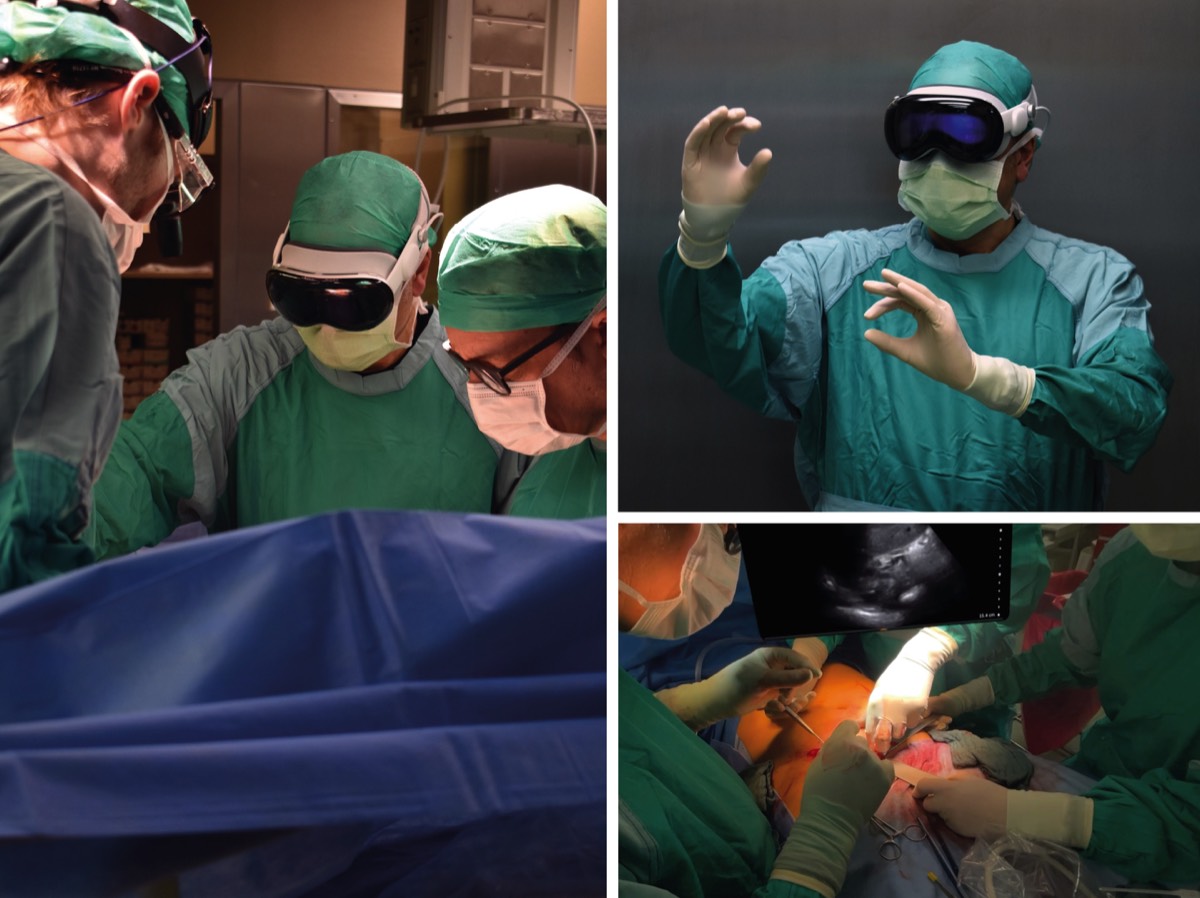
"Redefining the Laparoscopic Spatial Sense: AI-based Intra- and Postoperative Measurement from Stereoimages“ has been accepted for the 38th AAAI Conference on Artificial Intelligence and is available via https://doi.org/10.48550/arXiv.2311.09744. The publication is the result of a fruitful collaboration between Karlsruhe Institute of Technology (KIT), Fraunhofer FIT, University of Bayreuth, and Charité – Universitätsmedizin Berlin. Authors are Leopold Müller, Patrick Hemmer, Moritz Queisner, Igor Sauer, Simeon Allmendinger, Johannes Jakubik, Michael Vössing, and Niklas Kühl.

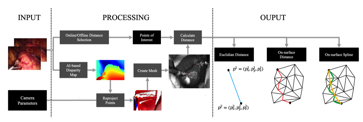
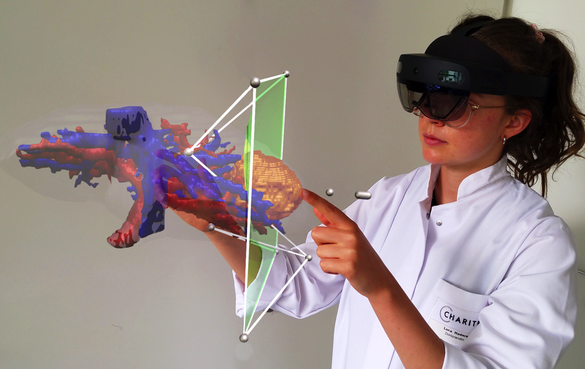
A significant challenge in image-guided surgery is the accurate measurement task of relevant structures such as vessel segments, resection margins, or bowel lengths. While this task is an essential component of many surgeries, it involves substantial human effort and is prone to inaccuracies. In this paper, we develop a novel human-AI-based method for laparoscopic measurements utilizing stereo vision that has been guided by practicing surgeons. Based on a holistic qualitative requirements analysis, this work proposes a comprehensive measurement method, which comprises state-of-the-art machine learning architectures, such as RAFT-Stereo and YOLOv8. The developed method is assessed in various realistic experimental evaluation environments. Our results outline the potential of our method achieving high accuracies in distance measurements with errors below 1 mm. Furthermore, on-surface measurements demonstrate robustness when applied in challenging environments with textureless regions. Overall, by addressing the inherent challenges of image-guided surgery, we lay the foundation for a more robust and accurate solution for intra- and postoperative measurements, enabling more precise, safe, and efficient surgical procedures.

Stacks Image 19612
Priv.-Doz. Dr. med. Karl Hillebrandt
Today Karl Hillebrandt gave an exzellent inaugural lecture entitled „Die Geister, die ich rief ... – Eine kurze Geschichte der De- und Rezellularisierung“ and is now a private lecturer (Privatdozent) at the Charité – Universitätsmedizin Berlin and habilitated in the field of "Experimental Surgery".

He is being honored for his achievements in the field of tissue engineering. His postdoctoral thesis is entitled "New approaches for the characterisation of decellularised tissues and the recellularisation of vessels".

Congratulations!

Stacks Image 19580
Rise of tissue- and species-specific 3D bioprinting based on decellularized extracellular matrix-derived bioinks and bioresins
Stacks Image 19562
The publication "Rise of tissue- and species-specific 3D bioprinting based on decellularized extracellular matrix-derived bioinks and bioresins" is now available online in Biomaterials and Biosystems. Authors are Laura Elomaa, Ahed Almalla, Eriselda Keshi, Karl H. Hillebrandt, Igor M. Sauer, and Marie Weinhart.

Thanks to its natural complexity and functionality, decellularized extracellular matrix (dECM) serves as an excellent foundation for creating highly cell-compatible bioinks and bioresins. This enables the bioprinted cells to thrive in an environment that closely mimics their native ECM composition and offers customizable biomechanical properties. To formulate dECM bioinks and bioresins, one must first pulverize and/or solubilize the dECM into non-crosslinked fragments, which can then be chemically modified as needed. In bioprinting, the solubilized dECM-derived material is typically deposited and/or crosslinked in a layer-by-layer fashion to build 3D hydrogel structures. Since the introduction of the first liver-derived dECM-based bioinks, a wide variety of decellularized tissue have been employed in bioprinting, including kidney, heart, cartilage, and adipose tissue among others. This review aims to summarize the critical steps involved in tissue-derived dECM bioprinting, starting from the decellularization of the ECM to the standardized formulation of bioinks and bioresins, ultimately leading to the reproducible bioprinting of tissue constructs.
New DFG research group FOR 5628 with our participation
Stacks Image 19539
The Deutsche Forschungsgemeinschaft (DFG, German Research Foundation) is establishing eight new research groups. One of these groups is FOR 5628: "Multiscale magnetic resonance elastography in cancer: The mechanical niche of tumor formation and metastatic spread – towards an improved diagnosis of cancer through mechanical imaging". The speaker and initiator is Prof. Ingolf Sack.

During the development of a tumour, the tissue changes its shape, e.g., alternating between hard and fluidic states. For this, cells exert forces and are simultaneously influenced by forces. This research group is investigating which mechanical-physical processes are behind this. How do tumours and metastases develop? What makes them resistant to therapy? The team is investigating these questions using magnetic resonance elastography (MRE) – a new clinical procedure that can be used to record the mechanical properties of body tissue. The goal is to be able to better diagnose tumours.
Dr. Karl Hillebrandt and Prof. Dr. Igor Sauer are part of the research group as PI in three projects:
  • A03 Cancer cell unjamming and jamming as prerequisites for the formation of primary and metastatic tumors
  • B03 Scaffold composition and fluid pressure in recellularized hepatic and pancreatic tumors
  • C01 Multiscale mechanical properties of tumors and tumor environment – from tissue specimens to patients

Was are happy to be part of this exzellent team!
Moderate LMWH anticoagulation improves success rate of hind limb allotransplantation in mice
Stacks Image 19589
The publication "Moderate LMWH Anticoagulation Improves Success Rate of Hind Limb Allotransplantation in Mice" is now available online in Plastic & Reconstructive Surgery-Global Open. Authors are B. Kern, M.-I. Ashraf, A. Reutzel-Selke, J. Mengwasser, D. Polenz, E. Michaels, J. Pratschke, S.G. Tullius, Ch. Witzel, and I.M. Sauer.

The mouse hind limb model represents a powerful research tool in vascularized composite tissue allotransplantation, but its applicability is limited due to poor graft survival (62%–83%). Vascular thrombosis and massive hemorrhage are the major causes for these drop-outs. We hypothesize that because of better anticoagulation effect and lower risk of thrombocytopenia, application of low molecular weight heparin (LMWH) will minimize vascular complications and enhance graft and animal survival.

Fifty allogeneic hind limb transplantations were performed (C57BL/6 to DBA/2 mice) using five different anticoagulation protocols. Bleeding and thromboembolic events were recorded macroscopically by postoperative hemorrhage and livid discoloration of the graft, respectively. Graft perfusion and survival were monitored daily by capillary-refill-time of graft toes within 2–3 seconds. Vascular congestion and tissue necrosis were examined by histological evaluation of hematoxylin-eosin-stained tissue sections.

All transplantations were technically successful. Increase in thromboembolic events and a concomitant decrease in bleeding events were observed with the decreasing concentration of heparin in the perfusion solution. Although treatment of donor and recipient with low dose of LMWH could not reduce thromboembolic events, moderate dose effectively reduced these events. Compared with the poor outcome of graft perfusion with heparin alone, additional treatment of donor and recipient with low dose of LMWH improved graft and animal survival by 18%. Interestingly, animals treated with moderate dose of LMWH demonstrated 100% graft and animal survival.
Treatment of donor and recipient mice with a moderate dose of LMWH prevents vascular complications and improves the outcome of murine hind limb transplants.
ECRT Consumable Grant - Advanced Scientists
Stacks Image 19460
Nils Haep successfully applied for funding from the ECRT Consumable Grant - Advanced Scientists. In his project, Nils is investigating the function of a cysteine-type endopeptidase and described mutations in the endopeptidase in induced pluripotent stem cell derived hepatocytes and their influence on Adiponutrin induced NAFLD.

Congratulations!
Priv.-Doz. Dr. med. Simon Moosburner
Today Simon Moosburner gave his inaugural lecture on "Liver Transplantation in Germany - Opportunities and Solutions for the Future". He is now – at the age of 28 (!) – a private lecturer (Privatdozent) at the Charité – Universitätsmedizin Berlin and habilitated in the field of "Experimental Surgery".

He is being honored for his achievements in the field of extracorporeal organ perfusion and organ transplantation. His postdoctoral thesis is entitled "Challenges and solutions in adults and children after liver transplantation".

Congratulations!

Stacks Image 19446
Dr. Zeynep Akbal
Stacks Image 19424
We are delighted to welcome Dr. Zeynep Akbal as a new member of the team!
Before she started as a post-doctoral researcher at the Digital Surgery Lab she studied communication sciences, media sciences, philosophy, and worked on developing her interdisciplinary method around virtual reality (VR) technology. She did her doctorate in philosophy at Universität Potsdam. Her dissertation is recently published as a monograph, titled "Lived-Body Experiences in Virtual Reality. A Phenomenology of the Virtual Body.”
Her research focuses on the intersection of philosophy of perception, cognitive sciences and VR. In her recent research project “Tactile Stimulation in VR” at Max Planck Institute for Human Cognitive and Brain Sciences, she focused on the behavioral consequences of haptic feedback in a VR task.

Wellcome to the team!
science x media Tandem Program: "From Slices to Spaces"
Prof. Dr. Moritz Queisner and Frédéric Eyl (Designer and Managing Director of TheGreenEyl) successfully applied to the Stiftung Charité for funding as a "science x media tandem".
The science x media tandems are the first programme in the new funding priority "Open Life Science". With this funding priority, the Charité Foundation is working to make the life sciences in Berlin more comprehensible and accessible to a broader public and to strengthen the trustworthiness of medical professionals.

Under the title "From Slices to Spaces", the tandem of Moritz Queisner and Frédéric Eyl is implementing a science parcours in which spatially complex research data from surgery and biomedicine will be made multisensually accessible to a broad audience through new visualization techniques. Building on research work on new imaging techniques by Moritz Queisner, they employ Extended Reality techniques. Due to their unique ability to link digital objects with the real environment of the viewers, the 4D images they generate are particularly suited for representing and conveying spatial information.

This is where the tandem's project comes in: 4D images are not only interesting for researchers to understand complex research data but can also provide laypeople with a less presupposing insight into research data and processes. Frédéric Eyl's media expertise will be used to make the specific visual knowledge from research comprehensible and experiential for non-experts. The science parcours is intended to integrate as a digital extension into the architecture of the new research building, "Der Simulierte Mensch", located on the premises of Charité. The parcours will include the facade, the inter-floor airspace, and the central glass surfaces within the building as its stations. By enabling users to explore 4D research data within the architecture and investigate it using their own smartphones in an AR application, concrete practices and deployment locations of new image-based technologies become experiential and comprehensible. This project not only enhances the perception of Charité and the scientific location of Berlin but also opens up places of knowledge creation to the public, making practices and techniques of life sciences more visible.


Stacks Image 15695
Eriselda Keshi and Simon Moosburner CSP fellows
Stacks Image 15700
Dr. Eriselda Keshi and Dr. Simon Moosburner successfully applied for the BIH Charité Clinician Scientist Program (CSP).
The program provides a unique opportunity for young medical doc- tors to combine their clinical training with protected time for research. This structured career path fosters translation of scientific discoveries into application and strengthens the innovative capacity of academic medicine.
Participants of the CSP devote 50 percent of their working hours to research over a period of three years.

Dr. Keshi will work on "NeoPancreasPrint, a 3D printed islet hosting tissue based on biocompatible ink derived from human decellularized pancreas".
Dr. Moosburner applied with this project "Alleviation of Senescence induced Ischemia-Reperfusion in Liver Grafts of Elderly Donors [SenEx".


Congratulations!
Prof. Dr. Nathanael Raschzok
Stacks Image 15709
Nathanael Raschzok | Experimental Surgery | 2007

With us in the team since he was a student, he has so far climbed all academic levels with flying colours.
In recognition of his outstanding achievements in research, teaching and the promotion of young academics, Nathanael was awarded the title of Associate Professor at the Charité – Universitätsmedizin Berlin.

Congratulations, Prof. Raschzok!
DFG | Grant for Machine Perfusion RCT
Stacks Image 19490
The German Research Foundation (Deutsche Forschungsgemeinschaft – DFG) s sponsoring a multicenter randomized controlled clinical trial by Prof. Dr. Georg Lurje entitled "End-ischemic hypothermic oxygenated (HOPE) or normothermic machine perfusion (NMP) compared to conventional cold storage (CCS) in donation after brain death (DBD) liver transplantation; a prospective multicenter randomized controlled trial (HOPE-NMP)".

The purpose of this study is to test the effects of end-ischemic NMP versus end-ischemic HOPE technique in a multicentre prospective randomized controlled clinical trial (RCT) on ECD liver grafts in DBD liver-transplantation (HOPE-NMP). Two-hundred-thirteen (n = 213) human whole organ liver grafts will be submitted to either NMP (n = 85) or HOPE (n = 85) directly before implantation and going to be compared to a control-group of patients (n = 43) transplanted with static cold storage preserved ECD-allografts. Primary (surgical complications as assessed by the comprehensive complication index [CCI]) and secondary (graft- and patient survival, hospital costs, hospital stay) endpoints are going to be analysed.


Congratulations !
Work with us | PhD position
We offer a funded PhD position for a computational biologist @ Experimental Surgery, Charité – Universitätsmedizin Berlin!

Are you interested to work on cutting-edge cancer research, and investigating complex-chromosomal as well as other genomic phenomena in human carcinoma cells ?

  • You will work on complex genome analyses (single-cell analyses, whole genome analyses) to detect, annotate and re-construct circular DNA using state of the art computational tools (e.g., Amplicon architect).
  • We provide motivation and high commitment in supervision in areas around experimental oncology and surgery.
  • The position allows the applicant to pursue an academic qualification, while collaborating and networking with international experts on extrachromosomal circular DNA and pancreatic carcinogenesis.

Apply now!
Send brief cover letter and CV via email to Dr. med. Matthäus Felsenstein (matthaeus.felsenstein@charite.de)
Dr. Eriselda Keshi
Stacks Image 15724
Today Eriselda Keshi successfully defended her doctoral thesis entitled "Construction of a thromboresistant vascular graft for vascular surgery purposes by decellularisation and recellularisation" summa cum laude!

Congratulations !
BIH Medical Student Research Stipend for Cao Zhong Jing Jin
Stacks Image 15729
Cao Zhong Jing Jin under the supervision of Dr. med. Matthäus Felsenstein successfully applied for the BIH Medical Student Research Stipend on their project “Deciphering the molecular determinants for the transformation of high-grade pancreatic duct dysplasia to invasive carcinoma by single-cell transcriptomics”. She is conducting a cutting-edge research project merging molecular data with histological information in collaboration with the BIH core facilities for single-cell genomics (Dr. Thomas Conrad) and intelligent imaging (Prof. Dr. Christian Conrad). Using modern single-cell- and spatial transcriptomics on pancreatic precursor and carcinoma samples, the project aims at defining molecular signatures that drive dysplastic cells.

Congratulations!
New DFG project "4D Imaging"
Stacks Image 15736
The DFG Schwerpunktprogramm „Das Digitale Bild“ (SPP 2172) funds the new project “4D Imaging: From Image Theory to Imaging Practice” (2023-2026). Principal investigators are Prof. Dr. Kathrin Friedrich (Universität Bonn) and Prof. Dr. Moritz Queisner.

The term 4D imaging refers to a new form of digital visuality in which image, action and space are inextricably interwoven. 4D technologies capture, process and transmit information about physical space and make it computable in real time. Changes due to movements and actions become calculable in real time, making 4D images particularly important in aesthetic and operational contexts where they reconceptualize various forms of human-computer interaction. The 4D Imaging project responds to the growing need in medicine to understand, use, and design these complex imaging techniques. It transfers critical reflexive knowledge from research into clinical practices to enable surgeons to use and apply 4D Imaging techniques. Especially in surgical planning, 4D Imaging techniques may improve the understanding and accessibility of spatially complex anatomical structures. To this end, the project is developing approaches to how 4D imaging can complement and transform established topographic ("2D") imaging practices.

Stacks Image 15738
Work with us | PhD position

We are hiring: 3-year #PhD position @Charité – Universitätsmedizin Berlin.
  • Join our interdisciplinary team for a PhD on new #imaging technologies at the intersection of digital health, surgery and biomedicine
  • Explore new ways to understand and/or visualize anatomical structures in #4D using extended reality #XR #digitaltransformation
  • Connect theory and practice in an interdisciplinary research group
  • Open call: open to all disciplines! Yes, that’s right – design, computer science, computer visualistics, digital health, psychology, media studies, workplace studies, game design…
  • What counts is a convincing idea for your doctoral project in the field of "4D imaging“

Sounds interesting? Apply now or reach out to Moritz Queisner (moritz.queisner@charite.de) if you have any questions.

More information:
German: https://karriere.charite.de/stellenangebote/detail/wissenschaftliche-mitarbeiterin-wissenschaftlicher-mitarbeiter-dwm-technologietransfer-chirurgie-dm27222a
English: https://karriere.charite.de/stellenangebote/detail/scientific-researcher-phd-position-dfm-dm27222b

EKFS grant for functional role and clinical relevance of ecDNA in pancreatic adenocarcinoma
Stacks Image 15749
Dr. Matthäus Felsenstein successfully applied for funding from the Else Kröner Fresenius Stiftung (EKFS) for his project “The functional role and clinical relevance of extrachromosomal DNA (ecDNA) in pancreatic adenocarcinoma”. In close collaboration with the excellence group around Professor Anton Henssen, he is aiming at improved understanding of unique genomic patterns as a results of complex chromosomal rearrangements in pancreatic adenocarcinoma that could drive its aggressive behavior. He will use state-of-the art three-dimensional tissue culture to enrich for neoplastic cells from primary PDAC specimen and subsequently perform genome analyses to identify samples that harbor extrachromosomal DNA. The clinical impact of these chromosomal structures will be explored by clinical correlation analyses and therapy response in vitro.

Congratulations!
"Einstein Kickbox - Advanced Scientists" grant
Stacks Image 15768
Nils Haep successfully applied for the "Einstein Kickbox - Advanced Scientists" grant of the Einstein Center for Regenerative Therapies (ECRT). The purpose of the grant is to provide start-up funding for interesting experimental projects in regenerative medicine.  In his project, Nils is investigating the function of a cysteine-type endopeptidase and described mutations in the endopeptidase in hepatocytes using live-cell imaging and metabolomics. Through this preliminary work, he hopes to generate a hypothesis on the function of the endopeptidase and the underlying mechanism of the mutations. In the next step, he plans to develop a disease model for fatty liver from induced pluripotent stem cells.
Design Lab #13: Material Legacies
The exhibition »Design Lab #13: Material Legacies« at Kunstgewerbemuseum Berlin, opening on November 3rd, 2022, explores contingencies and ruptures between traditional crafts and the most recent developments at the crossroads of material research, design, engineering, and architecture. It brings together artifacts from the museum’s collection with work-in-progress installations by designers and researchers from the Cluster of Excellence »Matters of Activity. Image Space Material« in order to initiate a dialogue about the historical, contemporary, and future conditions under which materiality unfolds.

By engaging with a series of different materials and techniques the exhibition encompasses both the problematization of unsustainable pasts and presents as well as the imagination of speculative material futures. Taking materiality as a starting point, each of the exhibits will investigate its sociocultural, economic, and political context in order to disentangle the multiple interrelations that arise from and with materials. As such »Design Lab #13: Material Legacies« aims to challenge the passive understandings of materiality and associate with the widening discourse on relational knowledge practices in arts, design, humanities, and social science.

The exhibition will be running from 4 November 2022 to 26 February 2023. For the exhibition announcement on the website of the
Staatliche Museen zu Berlin – Preußischer Kulturbesitz.

Stacks Image 15778


Exhibition Opening

3 November 2022, 6 pm

The opening event will include an introduction to the exhibition by Dr. Claudia Banz, Curator of Design at the Kunstgewerbemuseum Berlin, and Prof. Dr. Claudia Mareis, co-director of the Cluster of Excellence »Matters of Activity. Image Space Material«. Moreover, exhibition curators Michaela Büsse and Emile De Visscher will provide background on the exhibition, its goals, and how the curatorial process was undertaken.
The exhibition opening is part of the Berlin Science Week 2022.
Prof. Dr. Georg Lurje
Stacks Image 15787
In recognition of his exceptional achievements in research and teaching, Georg Lurje was honoured to receive an associate professorship in surgery at the Charité – Universitätsmedizin Berlin.

CONGRATULATIONS !
BMBF funds KIARA
With the programme "AI-based assistance systems for process-accompanying health applications", the Federal Ministry of Education and Research (BMBF) is funding innovative research and development work on interactive assistance systems that support processes in clinical health care using artificial intelligence methods.

Together with the partners Gebrüder Martin GmbH & Co. KG, Tuttlingen, HFC Human-Factors-Consult GmbH, Berlin and the Fraunhofer Institute for Telecommunications Heinrich-Hertz-Institut (HHI), Berlin, we successfully applied with the project "AI-based recording of work processes in the operating theatre for the automated compilation of the operating theatre report" (KIARA).


Stacks Image 15797


Operating theatre reports document all relevant information during surgical interventions. They serve to ensure therapeutic safety and accountability and as proof of performance. The preparation of the OR report is time-consuming and ties up valuable working time – time that is then not available for the treatment of patients.

In the KIARA project, we are working on a system that automatically drafts operating theatre reports. The KIARA system is intended to relieve medical staff: it documents operating theatre activities and creates a draft of the report, which then only needs to be checked, completed and approved. The system works via cameras integrated into operating theatre lamps. Their image data is then analysed with the help of artificial intelligence to recognise and record objects, people and all operating theatre activities. The ambitious system is to be developed and tested in a user-centred manner for procedures in the abdominal cavity and in oral and maxillofacial surgery.

KIARA is intended to continuously learn through human feedback and to simplify clinical processes for the benefit of medical staff by automating the creation of operating theatre reports. The system can also be applied to other operating theatre areas in the future.

The project has a financial volume of € 2.16 million.
The kick-off meeting took place on 16.09.2022 at the Charité.
„Si-M-Day“ | November 24th, 2022
Stacks Image 15807
Join us – at our online networking event.
We, the Si-M spokespersons and coordinators, are pleased to invite you to our first symposium „Si-M-Day“ on 24th November from 9 to 14 h – online.
It is dedicated to networking and initiation of projects between investigators of both partner institutions.
Click
here to register until November 18th (abstract submission deadline October 17th).
DFG Walter-Benjamin grant for the investigation of sex as a biological variable in alloimmunity
Stacks Image 15816
With a DFG Walter-Benjamin grant Dr. med. Friederike Martin will join the laboratory of Transplant Surgery Research at Harvard under the direction of Professor Stefan G. Tullius in Boston to investigate the role of biological sex for transplantation outcome.

Influences of donor and recipient sex on transplantation outcome have been described manifold, as well as an influence of sex hormones on the innate and adaptive immune response. So far, research, investigating the impact of sex hormones and different sex- and age-dependent sex-hormone levels on alloimmune response after solid organ transplantation is lacking. The aim of the project “Sex as a biological Variable in Alloimmunity” is, to delineate the impact of sex hormones and especially estrogens and age-dependent changes in estrogen-levels on alloimmune response after allogenic transplantation.  The project is based on the publication “Recipient sex and estradiol levels affect transplant outcomes in an age-specific fashion” published in the AJT in 2021 by the workgroup of Prof. Tullius.

Friederike, who already received the Sanofi Women in Transplantation fellowship grant for research in gender and sex in transplantation in 2021, will work as a Postdoc on this project in the Tullius Lab for an expected 2 years period, starting in January 2023.


Congratulations!
2022 TTS Mentee-Mentor-Award
Stacks Image 15822
Dr. Barbara Kern und Prof. Dr. Stefan G. Tullius received the 2022 Mentee-Mentor-Award of The Transplant Society during the 29th Conference in Buenos Aires.

In collaboration with National and International Societies, TTS acknowledges and recognizes the efforts of scientists who have advanced our understanding of transplantation science and fostered the development of young investigators.
The Mentee-Mentor Awards are designed to encourage dialogue and interactions between trainees and established investigators, and provide financial support for their joint participation in the Congress.

Congratulations!
Active Matter in Robotic-Assisted Surgery
Stacks Image 15834
Tuesday, 12.09.2022 | Cluster Retreat | Matters of Activity

2:30 – 2:45 pm Welcome & Intro
2:45 – 4:15 pm Panel 1
Rasa Weber Product Design (20 Minutes)
Felix Rasehorn Product Design (20 Minutes)
Binru Yang Engineering (20 Minutes)
Panel Discussion (30 Minutes)

4:15 – 4:45 pm Coffee Break
4:45 – 6:15 pm Panel 2
Jakub Rondomanski Mathematics (20 Minutes)
Babette Werner Art and Visual History (20 Minutes)
Anna Schäffner & Dominic Eger Domingos Product Design (20 Minutes)
Panel Discussion (30 Minutes)

6:15–7:30 pm Opening Exhibition und Aperitivo
Solid fraction determines stiffness and viscosity in decellularized pancreatic tissues
Stacks Image 15841
The article „Solid fraction determines stiffness and viscosity in decellularized pancreatic tissues“ in Biomaterials Advances is now available online.
There is free access to a PDF of the article here until August 20, 2022!

The role of extracellular matrix (ECM) composition and turnover in mechano-signaling and the metamorphic fate of cells seeded into decellularized tissue can be elucidated by recent developments in non-invasive imaging and biotechnological analysis methods. Because these methods allow accurate quantification of the composition and structural integrity of the ECM, they can be critical in establishing standardized decellularization protocols. This study proposes quantification of the solid fraction, the single-component fraction and the viscoelasticity of decellularized pancreatic tissues using compact multifrequency magnetic resonance elastography (MRE) to assess the efficiency and quality of decellularization protocols. MRE of native and decellularized pancreatic tissues showed that viscoelasticity parameters depend according to a power law on the solid fraction of the decellularized matrix. The parameters can thus be used as highly sensitive markers of the mechanical integrity of soft tissues. Compact MRE allows consistent and noninvasive quantification of the viscoelastic properties of decellularized tissue. Such a method is urgently needed for the standardized monitoring of decellularization processes, evaluation of mechanical ECM properties, and quantification of the integrity of solid structural elements remaining in the decellularized tissue matrix.

Authors are Joachim Snellings, Eriselda Keshi, Peter Tang, Assal Daneshgar, Esther C. Willma, Luna Haderer, Oliver Klein, Felix Krenzien, Thomas Malink, Patrick Asbach, Johann Pratschke, Igor M. Sauer, Jürgen Braun, Ingolf Sack, and Karl Hillebrandt.

Inaugural Lectures
We are pleased to announce that four members of staff have successfully completed their habilitation work in the last few months!

On
Friday, 08.07.2022 at 15:00 in lecture hall 3 of the teaching building (Forum 3, CVK), Dr. med. habil. Linda Feldbrügge and Dr. med. habil. Paul Ritschl will give their inaugural lectures entitled "New role of surgery in modern tumour and transplant medicine".

On
Friday, 15.07.2022 at 16:30 in the Friedrich Kopsch lecture theatre of the Anatomy Department at Campus Mitte Dr. med. habil. Eva Dobrindt and Dr. med. habil. Rosa Schmuck will present their inaugural lectures with the topic "An Operating Room of One's Own - The Surgeon in Ancient Tradition and Modernity".
This will be followed by a small reception in the park in front of the venue.
Stacks Image 15851
Si-M | Topping-out Ceremony
Today, representatives of Charité – Universitätsmedizin Berlin and Technische Universität Berlin celebrated the topping-out ceremony for the research building "Der Simulierte Mensch" (Si-M, "The Simulated Human") together with political representatives. Guests included the Governing Mayor Franziska Giffey, Senator for Health and Science and Charité Supervisory Board Chair Ulrike Gote and Finance Senator Daniel Wesener.

We are very excited: this will be a great building with even greater content.

Stacks Image 15861
VolumetricOR | Surgical Innovation
Stacks Image 15866
Our paper "VolumetricOR: A new Approach to Simulate Surgical Interventions in Virtual Reality for Training and Education" is available in the latest issue of Surgical Innovation.

Surgical training is primarily carried out through observation during assistance or on-site classes, by watching videos as well as by different formats of simulation. The simulation of physical presence in the operating theatre in virtual reality might complement these necessary experiences. A prerequisite is a new education concept for virtual classes that communicates the unique workflows and decision-making paths of surgical health professions (i.e. surgeons, anesthesiologists, and surgical assistants) in an authentic and immersive way. For this project, media scientists, designers and surgeons worked together to develop the foundations for new ways of conveying knowledge using virtual reality in surgery.
A technical workflow to record and present volumetric videos of surgical interventions in a photorealistic virtual operating room was developed. Situated in the virtual reality demonstrator called VolumetricOR, users can experience and navigate through surgical workflows as if they are physically present . The concept is compared with traditional video-based formats of digital simulation in surgical training.

VolumetricOR let trainees experience surgical action and workflows a) three-dimensionally, b) from any perspective and c) in real scale. This improves the linking of theoretical expertise and practical application of knowledge and shifts the learning experience from observation to participation.
Discussion: Volumetric training environments allow trainees to acquire procedural knowledge before going to the operating room and could improve the efficiency and quality of the learning and training process for professional staff by communicating techniques and workflows when the possibilities of training on-site are limited.

Authors are Moritz Queisner, Michael Pogorzhelskiy, Christopher Remde, Johann Pratschke, and Igor M. Sauer.
Tissue Engineering for the Diaphragm
"Tissue Engineering for the Diaphragm and its Various Therapeutic Possibilities – A Systematic Review" is available here in Advanced Therapeutics (open access).

Diaphragmatic impairments exhibit high morbidity as well as mortality while current treatment options remain unsatisfactory. Tissue engineering (TE) approaches have explored the generation of an optimal biocompatible scaffold for diaphragmatic repair through tissue decellularization or de novo construction, with or without the addition of cells. The authors conducted a systematic review on the current state of the art in diaphragmatic tissue engineering (DTE) and found 24 articles eligible for final synthesis. The included approaches studied decellularization-based graft generation and de novo bioscaffold construction. Three studies focused on in vitro host-scaffold interaction with synthesized, recellularized grafts and decellularized extracellular matrix scaffolds. Another three studies investigated evaluation tools for decellularization efficacy. Among all studies, recellularization is performed in both decellularization-based and de novo generated scaffolds. De novo constructed biocomposites as well as decellularized and recellularized scaffolds induced pro-regenerative remodeling and recovery of diaphragmatic function in all examined animal models. Potential therapeutic applications comprise substance defects requiring patch repair, such as congenital diaphragmatic hernia, and functional diseases demanding an entire organ transplant, like muscular dystrophies or dysfunction after prolonged artificial respiration.

Autors are Agnes K. Boehm, Karl H. Hillebrandt, Tomasz Dziodzio, Felix Krenzien, Jens Neudecker, Simone Spuler, Johann Pratschke, Igor M. Sauer, and Marco N. Andreas.
Stacks Image 15876
Detection of nicotinamide adenine dinucleotide (NAD) in cells and blood plasma
Stacks Image 15881
Priv,-Doz. Dr. med. Felix Krenzien and Dr. Jennifer Kirwan (Technologieplattform Metabolomik, Max-Delbrück-Centrum für Molekulare Medizin, Berlin) successfully applied for a grant within the Else Kröner Fresenius Stiftung funding line: Translational Research.

Recently, the molecule nicotinamide adenine dinucleotide (NAD) has attracted attention as it is involved in various important regulatory mechanisms, immune signaling, aging and regenerative processes. In this regard, it occupies key positions in many redox reactions of the body due to its role as a redox couple (NAD as an oxidized species and NADH as a reduced species). Consequently, NAD homeostasis (the maintenance of NAD in cells) is considered essential. The scientific consensus for many years was that the oxidized species resides exclusively in the intracellular milieu (iNAD). However, recent findings indicate that NAD also exists extracellularly (eNAD) and it is present in virtually all body fluids (from lymph to saliva to blood plasma). Based on these findings, precursors of NAD have recently been approved by the FDA and are commercially available. Measurement of eNAD in blood plasma is problematic due to its low concentration in the nanomolar range. However, quantifying eNAD plasma levels but also eNAD concentrations in cells is necessary to monitor the intake of NAD or its precursors and to adjust their dosage precisely.
The primary objective of this project is to validate, bioanalyze,and to document the assay for eNAD according to the ICH-M10 guidance document endorsed by the U. S. Food and Drug Administration (FDA). Adherence to the principles presented in this guideline should improve the quality and consistency of bioanalytical data, thereby supporting assay development and market approval. In addition, the assay will also be established for the measurement of intracellular NAD (iNAD), and validation of iNAD quantification will also be performed according to the guideline.

In the second part of the project, a clinical study will be conducted to determine whether the intake of nicotinamide riboside (precursor of NAD) leads to a change in eNAD and iNAD. Thus, the basis for an indication-dependent bioanalysis of the measurement of NAD will be developed to monitor the intake of NAD and its precursor or to adjust the dosage specifically on the basis of the quantification.
Stacks Image 15883

DFG Funds Extension of the Digital Clinician Scientist Program
Stacks Image 15892
Following the successful application for approval of the continuation of the BIH Charité Digital Clinician Scientist Program (DCSP) by the DFG, additional funding of around 1.3 million euros is now available over a period of two years: The DFG funding benefits physicians at Charité who have embarked on a scientific medical career path and, with their innovative research projects, are already playing a key role in shaping the digital transformation of healthcare during their residency training.

The Digital Clinician Scientist Program (DCSP) was jointly initiated by the German Research Foundation (DFG), the BIH, and the faculty of Charité - Universitätsmedizin Berlin in early 2019. The DCSP is an extension of the successful BIH Charité Clinician Scientist Program, which has set standards in the medical research landscape throughout Germany. The structured career path enables researching physicians to build the foundation for a successful career as a clinician scientist by providing protected time for research activities and non-clinical training during their residency. The DCSP is intended for clinically active physicians who are already actively shaping the digital transformation process of healthcare with their innovative research projects during their residency training. The main applicant of the continuation application is Prof. Dr. Igor M. Sauer, Director of the BIH Charité Digital Clinician Scientist Program, Deputy Clinic Director of the Department of Surgery, and Head of the Experimental Surgery at Charité. Since the start of the program in 2019, 24 physicians have benefited from funding, thus a broad spectrum of digital topics is already being addressed in various clinics at the Charité. The DFG originally funded the program for three years with more than three million euros. With the approved extension, the funding program now has a further 1.3 million euros available over a period of two years.
Development of human-based hydrogels as a substitute for mouse-derived Matrigel for cancer research
Stacks Image 15897
With a new funding line, Charité 3R wants to support the development of animal-free cell cultures at Charité.

For in vitro cancer research, mini-tumours are grown in a gel-like cultivation structure that serves the three-dimensional growth of the mini-tumours. This gel-like cultivation substance is obtained from mouse tumours, an unnatural cultivation environment for human mini-tumours. The aim of the project "Development of human-based hydrogels as a substitute for mouse-derived Matrigel for cancer research" by Björn Papke from the Institute of Pathology and Karl Hillebrandt is to produce a cultivation structure without animal additives. For this purpose, a cultivation structure, also gel-like, is to be produced from tissue obtained from patients during surgical procedures, which better corresponds to the natural environment of the human mini-tumours.


Congratulations!
AI-based Risk Assessment in Pancreatic Surgery
Stacks Image 15902
Our work on „Perioperative Risk Assessment in Pancreatic Surgery Using Machine Learning“ was published on the occasion of the 43rd Annual International Conference of the IEEE Engineering in Medicine and Biology Society (EMBC).

Pancreatic surgery is associated with a high risk for postoperative complications and death of patients. Complications occur in a variable interval after the procedure. Often, a patient has already left the ICU and is not properly monitored anymore when the complication occurs. Risk stratification models can assist in identifying patients at risk in order to keep these patients in ICU for longer. This, in turn, helps to identify complications earlier and increase survival rates. We trained multiple machine learning models on pre-, intra- and short term postoperative data from patients who underwent pancreatic resection at the Department of Surgery, Campus Charité Mitte | Campus Virchow-Klinikum, Charité - Universitätsmedizin Berlin. The presented models achieve an area under the precision-recall curve (AUPRC) of up to 0.51 for predicting patient death and 0.53 for predicting a specific major complication. Overall, we found that a classical logistic regression model performs best for the investigated classification tasks. As more patient data becomes available throughout the perioperative stay, the performance of the risk stratification model improves and should therefore repeatedly be computed.

Authors are Bjarne Pfitzner, Jonas Chromik, Rachel Brabender, Eric Fischer, Alexander Kromer, Axel Winter, Simon Moosburner, Igor M. Sauer, Thomas Malinka, Johann Pratschke, Bert Arnrich, and Max M. Maurer.
Annu Int Conf IEEE Eng Med Biol Soc. 2021 Nov;2021:2211-2214. doi: 10.1109/EMBC46164.2021.9630897.
Dr. med. Hannah Everwien
Stacks Image 15907
Hannah Everwien successfully defended her doctoral thesis entitled "Construction of a neo-pancreas by means of decellularisation and recellularisation" (summa cum laude)!

Congratulations!

Position for Student Assistant
Stacks Image 15912
Project: This project aims to analyse purinergic signal pathways, especially ecto-enzymes of the CD39 family and their role in tumor immunology. Tissue expression as well as the activity of the associated purinergic molecules (receptors, signaling molecules and degrading enzymes) will be examined in a patient cohort of different gastrointestinal tumors with and without (peritoneal) metastases.

Methods: Clinical tissue acquisition (perioperative of patients undergoing surgery), FACS, immunohistology, qPCR, cell culture.

Requirements: Medical student with clinical experience with patients (i.e. "Famulaturen") and drawing blood. Above average scientific interest and engagement. Teamwork and self sufficiency.

What we offer: Learning of scientific methods in an excellently equipped laboratory, teamwork. Mentoring in the clinical and lab. Publication of results and potential of a doctoral thesis.

Data: 10h/week. Project duration: 9 months. Begin: November 2021 (flexible). Salary according to TV Stud III for Berlin.

If you're the right person: please send all application documents, e.g. cover letter, curriculum vitae, certificates, attestations, etc. to the following address, quoting the reference number by e-mail to
Charité – Universitätsmedizin Berlin
Chirurgische Klinik, Exp. Chirurgie
z.Hd. Dr. Linda Feldbrügge
Augustenburger Platz 1
D-13353 Berlin
linda.feldbruegge@charite.de
BMBF grant – GreifbAR
Stacks Image 15917
The Federal Ministry of Education and Research (BMBF) funds the project "Tangible reality - skilful interaction of user hands and fingers with real tools in mixed reality worlds (GreifbAR)" – a cooperation of the Augmented Vision group of the DFKI (Prof. Dr. Didier Stricker), the Department of Psychology and Human-Machine Interaction of the University of Passau (Prof. Dr. Susanne Mayr), the company NMY Mixed Reality Communication (Christoph Lenk), and the Experimental Surgery of Charité – Universitätsmedizin Berlin (Prof. Dr. Igor M. Sauer).

The goal of the GreifbAR project is to make extended reality (XR) worlds, including virtual (VR) and mixed reality (MR), tangible and graspable by allowing users to interact with real and virtual objects with their bare hands. Hand accuracy and dexterity is paramount for performing precise tasks in many fields, but the capture of hand-object interaction in current XR systems is woefully inadequate. Current systems rely on hand-held controllers or capture devices that are limited to hand gestures without contact with real objects. GreifbAR solves this limitation by proposing a sensing system that detects both the full hand grip including hand surface and object pose when users interact with real objects or tools. This sensing system will be integrated into a mixed reality training simulator.

Competent handling of instruments and suture material is the basis of every surgical activity. The main instruments used in surgery are in the hands of the surgical staff. Their work is characterised by the targeted use of a large number of instruments that have to be operated and controlled in different ways. Until now, surgical knotting techniques have been learned by means of personal instruction by experienced surgeons, blackboard images and video-based tutorials. A training and teaching concept based on the acquisition of finger movement does not yet exist in surgical education and training. Learning surgical account techniques through participant observation and direct instruction by experienced surgeons is cost-intensive and hardly scalable. This type of training is increasingly reaching its limits in daily clinical practice, which can be attributed in particular to the changed economic, social and regulatory conditions in surgical practice. Students and trainees as well as specialist staff in further training are therefore faced with the problem of applying and practising acquired theoretical knowledge in a practice-oriented manner. Text- and image-based media allow scalable theoretical knowledge acquisition independent of time and place. However, gestures and work steps can only be passively observed and subsequently imitated. Moreover, the learning success cannot be quantitatively measured and verified.

The aim of the Charité's sub-project is therefore to develop a surgical application scenario for Mixed/Augmented Reality (MR/AR) for the spatial guidance and verifying recording of complex fine motor finger movements for the creation of surgical knots, the practical implementation and technical testing of the developed concept within the framework of a demonstrator, and the evaluation of the usability of the system for use in a clinical context.
Two new research grants by Berliner Krebsgesellschaft
Stacks Image 15922
The Berliner Krebsgesellschaft will fund two very interesting research projects by Dr. Linda Feldbrügge and Dr. Karl Hillebrandt in collaboration with Dr. Björn Papke.

Stacks Image 15929
„Purinergic immune regulation in peritoneal metastases of gastric cancer via CD39 and ENTPD3 – target for a novel immune Checkpoint inhibition?“ – PI: Dr. Linda Feldbrügge

Peritoneal metastasis, especially derived from gastric cancer (GC), has a poor prognosis with a median survival of only months. Treatment is usually confined to palliative systemic chemotherapy, complemented individually by checkpoint inhibitors that block PD1-signaling. Innovative therapies combining surgery with local drug application such as hyperthermic intraperitoneal chemotherapy (HIPEC) or pressurized intraperitoneal aerosol chemotherapy (PIPAC) are still pending confirmation in clinical trials. Purinergic signaling, which involves ATP hydrolysis and generation of adenosine, regulated through CD39 (ENTPD1) and related enzymes, has been recognized as a critical immunoregulatory pathway in the tumor microenvironment (TME). The objective of the current project is to characterize the immune environment in the unique setting of peritoneal metastasis of gastric cancer with a focus on ectonucleotidases CD39 and ENTPD3 on T cells, macrophages and MDSC as well as mechanisms of ectonucleotidase-mediated immune regulation in tumor associated macrophages in vitro. As a high-volume center for surgical therapy of peritoneal malignancies and with years of experience in ectonucleotidase research, we aim to advance the understanding of peritoneal metastasis and contribute to improving treatment options for our patients.

Stacks Image 15936
"The influence of decellularised tumour matrix heterogeneity in relation to KRAS/MAPK inhibition of in vitro colorectal liver metastases." PI: Dr. Karl Hillebrandt and Dr. Björn Papke (Dept. of Pathology)

Colorectal cancer (CRC) is the second leading cause of cancer-related death worldwide, with approximately 900,000 annual deaths. 30-50% of patients develop colorectal liver metastases (CRLM) during their disease. More than 50% of these tumors have mutations in the KRAS oncogene, making them usually poorly treatable. Despite multimodal therapy concepts have improved the outcome of these patients, a large proportion of patients suffer a recurrence of their disease. For better therapeutic concepts, we need to better understand the tumor biology and metastatic mechanisms of these diseases. In vitro models, such as two-dimensional cell culture, are primarily used for this purpose. These models can only reflect the physiological complexity to a limited extent. Recently, it was shown that the use of organ-specific and tumor-specific extracellular matrix (ECM) has an impact on the behavior of human CRC cell lines. Culture of cell lines with decellularized matrix resulted in cells adopting a metastatic cell state and forming significantly more metastases in a mouse model than cells cultured on plastic or collagen. The goal of our project is to study the growth (with and without inhibition of the RAS/MAPK signaling pathway) of patient-derived tumor organoids growing on different decellularized metastatic matrices (dMM) and decellularized liver matrices (dLM). These studies of tumor matrix heterogeneity are essential to define which starting materials, for in vitro modeling of our three-dimensional tumor organoid culture, can be used to develop the most physiological, personalized dLM/dMM-based CRLM in vitro model possible. Based on these results, we plan to conduct small-scale therapy evaluations for personalized tumor therapy using our in vitro dLM/dMM-based CRLM in the near future.

Congratulations!
 Page 1 / 6  >>

Archive


Categories

Year

This website or its third-party tools use cookies, which are necessary to its functioning and required to achieve the purpose illustrated in the Disclaimer. By closing this banner, scrolling this page, clicking a link or continuing to browse otherwise, you agree to the use of cookies.