News

Impact of Percoll purification on isolation of primary human hepatocytes
Stacks Image 16198
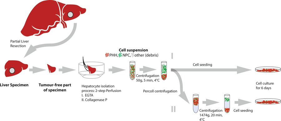
Scientific Reports published our paper "Impact of Percoll purification on isolation of primary human hepatocytes" (Sci Rep. 2019 Apr 25;9(1):6542). Authors are R. Horner, J.G.M.V. Gassner, M. Kluge, P. Tang, S. Lippert, K.H. Hillebrandt, S. Moosburner, A. Reutzel-Selke, J. Pratschke, I.M. Sauer, and N. Raschzok.

Research and therapeutic applications create a high demand for primary human hepatocytes. The limiting factor for their utilization is the availability of metabolically active hepatocytes in large quantities. Centrifugation through Percoll, which is commonly performed during hepatocyte isolation, has so far not been systematically evaluated in the scientific literature. 27 hepatocyte isolations were performed using a two-step perfusion technique on tissue obtained from partial liver resections. Cells were seeded with or without having undergone the centrifugation step through 25% Percoll. Cell yield, function, purity, viability and rate of bacterial contamination were assessed over a period of 6 days. Viable yield without Percoll purification was 42.4 × 106 (SEM ± 4.6 × 106) cells/g tissue. An average of 59% of cells were recovered after Percoll treatment. There were neither significant differences in the functional performance of cells, nor regarding presence of non-parenchymal liver cells. In five cases with initial viability of <80%, viability was significantly increased by Percoll purification (71.6 to 87.7%, p = 0.03). Considering our data and the massive cell loss due to Percoll purification, we suggest that this step can be omitted if the initial viability is high, whereas low viabilities can be improved by Percoll centrifugation.
Isolation of primary human hepatocytes & LiMax-test
Stacks Image 16416
Tissue Engineering (Part C: Methods) accepted our paper entitled "The predictive value of the LiMAx-test for the isolation of primary human hepatocytes".
Authors are R.D. Major, M. Kluge, M. Jara, M. Nösser, R. Horner, J. Gassner, B. Struecker, P. Tang, S. Lippert, A. Reutzel-Selke, D. Geisel, T. Denecke, M. Stockmann, J. Pratschke, I.M. Sauer, and N. Raschzok.

The need for primary human hepatocytes is constantly growing, for basic research as well as for therapeutic applications. However, the isolation outcome strongly depends on the quality of liver tissue, and we are still lacking a preoperative test that allows the prediction of the hepatocyte isolation outcome. Here we evaluated the “maximal liver function capacity test” (LiMAx) as predictive test for the quantitative and qualitative outcome of hepatocyte isolation. This test is already used in clinical routine to measure preoperative and to predict postoperative liver function.
The patient’s preoperative mean LiMAx was obtained from the patient records and preoperative CT and MRI images were used to calculate the whole liver volume in order to adjust the mean LiMAx. The outcome parameters of the hepatocyte isolation procedures were analyzed in correlation with the adjusted mean LiMAx.
Primary human hepatocytes were isolated from partial hepatectomies (n=64).
From these 64 hepatectomies we included 48 to our study and correlated their isolation outcome parameters with volume corrected LiMAx values. From a total of 11 hepatocyte isolation procedures, metabolic parameters (albumin, urea and aspartate aminotransferase) were assessed during the hepatocyte cultivation period of 5 days. The volume adjusted mean LiMAx showed a significant positive correlation with the total cell yield (p= 0.049;r= 0.242;n= 48). The correlations of volume adjusted LiMAx values with viable cell yield and cell viability did not reach statistical significance. A sub-group analysis of isolations from patients with colorectal metastasis revealed a significant correlation between volume adjusted mean LiMAx and total cell yield (p= 0.012;r= 0.488;n= 21) and viable cell yield (p=0.034;r=0.405;n=21). Whereas a sub-group analysis of isolations of patients with carcinoma of the biliary tree showed significant correlations of volume adjusted mean LiMAx with cell viability (r= 0.387;p= 0.046;n=20) and lacked significant correlations with total cell yield (r= - 0.060;p= 0.401;n=20) and viable cell yield (r= 0.012;p= 0.480;n=20). The volume-adjusted mean LiMAx did not show a significant correlation with any of the metabolic parameters. In conclusion, the LiMAx-test might be a useful tool to predict the quantitative outcome of hepatocyte isolation, as long as underlying liver disease is taken into consideration.
Read More
Cells isolated from diseased explanted livers
Stacks Image 16496
The International Journal of Artificial Organs (official journal of the European Society for Artificial Organs [ESAO]) published our paper on Isolation, characterization and cold storage of cells isolated from diseased explanted livers. Authors are Belaschk E, Rohn S, Mukiibi R, Reutzel-Selke A, Tang P, Sawitzki B, Pratschke J, Sauer IM and Mogl MT.

Livers discarded after standard organ retrieval are commonly used as a cell source for hepatocyte transplantation. Due to the scarcity of organ donors, this leads to a shortage of suitable cells for transplantation. Here, the isolation of liver cells from diseased livers removed during liver transplantation is studied and compared to the isolation of cells from liver specimens obtained during partial liver resection. Hepatocytes from 20 diseased explanted livers (Ex-group) were isolated, cultured and stored at 4°C for up to 48 hours, and compared to hepatocytes isolated from the normal liver tissue of 14 liver lobe resections (Rx-group). The nonparenchymal cell fraction (NPC) was analyzed by flow cytometry to identify potential liver progenitor cells, and OptiPrep™ (Sigma-Aldrich) density gradient centrifugation was used to enrich the progenitor cells for immediate transplantation. There were no differences in viability, cell integrity and metabolic activity in cell culture and survival after cold storage when comparing the hepatocytes from the Rx-group and the Ex-group. In some cases, the latter group showed tendencies of increased resistance to isolation and storage procedures. The NPC of the Ex-group livers contained considerably more EpCAM+ and significantly more CD90+ cells than the Rx-group. Progenitor cell enrichment was not sufficient for clinical application. Hepatocytes isolated from diseased explanted livers showed the essential characteristics of being adequate for cell transplantation. Increased numbers of liver progenitor cells can be isolated from diseased explanted livers. These results support the feasibility of using diseased explanted livers as a cell source for liver cell transplantation.
Read More
Hepatocyte isolation after laparoscopic liver resection
Stacks Image 16675
Tissue Engineering, Part C: Methods accepted our paper entitled "Hepatocyte isolation after laparoscopic liver resection" for publication. Authors are Horner R*, Kluge M*, Gassner J, Nösser M, Major RD, Reutzel-Selke A, Leder AK, Struecker B, Morgul MH, Pratschke J, Sauer IM, Raschzok N (*contributed equally).

Liver tissue obtained from partial hepatectomy is a common source for isolation of primary human hepatocytes. Until now, liver resections were most commonly performed by conventional open surgery. Although the laparoscopic approach is currently emerging in liver surgery, data on the outcome of hepatocyte isolation from laparoscopically resected liver tissue is not available. A total of 22 hepatocyte isolations were performed using the two-step collagenase perfusion technique from October 2015 until March 2016. Liver tissue was obtained from n=15 open liver resections (OLR) and n=7 laparoscopic liver resections (LLR). Isolation parameters (cell yield, viability, percoll survival) were assessed and hepatocyte function (plating efficiency, urea, albumin, and aspartate aminotransferase) was measured over a culture period of 6 days (OLR: n=13; LLR: n=3). Total cell yield (OLR: 36.81 ± 6.77 x10⁶ cells/g vs. LLR 16.84 ± 10.66 x10⁶ cells/g, p=0.0318) as well as viable yield (OLR 31.70 ± 6.05 x10⁶ cells/g vs. LLL 14.70 ± 9.89 x10⁶ cells/g, p=0.0260) were significantly higher in the OLR group. Subgroup analysis revealed that the worse outcome of isolation of laparoscopically resected liver tissue was associated with right-lateral laparoscopic liver resections, while hepatocyte isolation from left-lateral laparoscopic liver resections was as effective as from open surgery. Hepatocyte function did not differ between hepatocytes from openly resected versus left-lateral laparoscopically resected liver tissue. We here present the first data on hepatocyte isolation from laparoscopic liver surgery. While the overall outcome is worse compared to open surgery, our data suggest that liver tissue from laparoscopic resection of the left lobe is an excellent source for primary human hepatocytes.
Read More
 Page 1 / 1 

Archive


Categories

Year

This website or its third-party tools use cookies, which are necessary to its functioning and required to achieve the purpose illustrated in the Disclaimer. By closing this banner, scrolling this page, clicking a link or continuing to browse otherwise, you agree to the use of cookies.