News

SPARK Berlin supports Fikatas Knot
Stacks Image 16694
The Berlin Institute of Health (BIH) and Stiftung Charité teamed up with Stanford University School of Medicine to initiate SPARK Berlin.

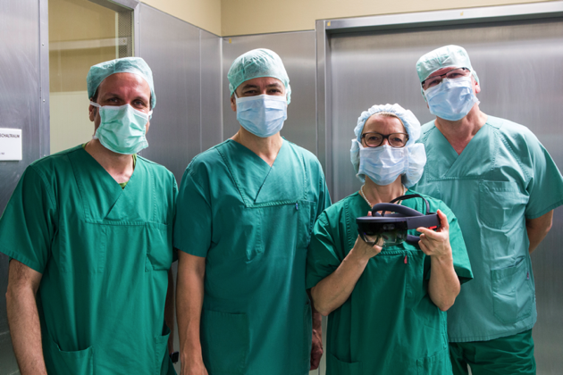
We are very pleased to announce that Dr. Panagiotis Fikatas' project “Device for ready-prepared surgical knots” was selected for both, funding and mentorship. 

SPARK was created to overcome the hurdles associated with translating academic discoveries into therapeutics and diagnostics that address unmet medical needs.  The SPARK mission is to help academics overcome the obstacles involved in moving their early discoveries from bench to bedside, to educate faculty, postdoctoral fellows and graduate students on the translational research process and path to clinical application, so that development of promising discoveries becomes second nature to our institution, and to develop more cost-effective and innovative approaches to drug development .

Congratulations!
Read More
Einladunfgzur öffentlichen Abschlusspräsentation
Stacks Image 17125
Sehr geehrte Damen und Herren,
Dear ladies and gentlemen,

Sie sind herzlich eingeladen, unserer öffentlichen Abschlusspräsentation unseres EU/EFRE-Projekts am 20.03.2013 um 15:00 Uhr beizuwohnen. Zusammen mit unseren Partnern der Firma microparticles GmbH werden wir den aktuellen Stand zur „Entwicklung von Partikel zur Detektion und ultralokoregionären Stimulation transplantierter Leberzellen“ darlegen.
You are cordially invited to attend our public presentation of our EU/EFRE project. Together with our partners of microparticles GmbH we will present our latest results concerning the „Development of particles for detection and ultralocoregional stimulation of transplanted liver cells“.

Wann/When?
20.03.2013 um/at 15:00

Wo/Where?
Experimentelle Chirurgie und Regenerative Medizin
Klinik für Allgemein-, Viszeral- und Transplantationschirurgie
Charité, Campus Virchow-Klinikum
Forschungshaus/BMFZ
Pilzraum, 1.OG

Wir wären dankbar, wenn Sie Ihr Kommen via email (anja.selke@charite.de)bestätigen könnten.
RSVP via email (anja.selke@charite.de).
Read More
CARS microscopy of MPIO
Stacks Image 17285
Following a successful project sponsored by the BMBF G. Pless, I.M. Sauer and U. Rauen report on the "Improvement of the cold storage of isolated human hepatocytes" (Cell Transplant. 2011 Jun 7. [Epub ahead of print]).
Increasing amounts of human hepatocytes are needed for clinical applications and different fields of research, such as cell transplantation, bioartificial liver support and pharmacological testing. This demand calls for adequate storage options for isolated human liver cells. As cryopreservation results in severe cryoinjury, short term storage is currently performed at 2-8º C in preservation solutions developed for the storage of solid organs. However, besides slowing down cell metabolism, cold also induces cell injury, which is, in many cell types, iron-dependent and not counteracted by current storage solutions. In this study, we aimed to characterize storage injury to human hepatocytes and develop a customized solution for cold storage of these cells. Human hepatocytes were isolated from material obtained from partial liver resections, seeded in monolayer cultures and, after a pre-culture period, stored in the cold in classical and new solutions followed by rewarming in cell culture medium.Human hepatocytes displayed cold-induced injury, resulting in > 80% cell death (LDH release) after one week of cold storage in University of Wisconsin solution or cell culture medium and 3 h of rewarming. Cold-induced injury could be significantly reduced by the addition of the iron chelators deferoxamine and LK 614. Experiments with modified solutions based on the new organ preservation solution Custodiol-N showed that ion-rich variants were better than ion-poor variants, chloride-rich solutions better than chloride-poor solutions, potassium as main cation superior to sodium and pH 7.0 superior to pH 7.4. LDH release after two weeks of cold storage in the thus optimized solution was below 20%, greatly improving cold storage of human hepatocytes. The results were confirmed by the assessment of hepatocellular mitochondrial membrane potential and functional parameters (resazurin reduction, glucacon-stimulated glucose liberation) and thus suggest the use of a customized hepatocyte storage solution for the cold storage of these cells.
Read More
Improved cold storage of human hepatocytes
Stacks Image 17315
As a first result of our latest projects concerning the role of miRNA in liver regeneration the American Journal of Physiology - Regulatory, Integrative and Comparative Physiology has accepted our paper "Temporal expression profiles indicate a primary function for microRNA during the peak of DNA replication after rat partial hepatectomy": The liver has the unique capacity to regenerate after surgical resection. However, the regulation of liver regeneration is not completely understood. Recent reports indicate an essential role for small non-coding microRNAs (miRNAs) in the regulation of hepatic development, carcinogenesis, and early regeneration. We hypothesized that miRNAs are critically involved in all phases of liver regeneration after partial hepatectomy. We performed miRNA microarray analyses after 70% partial hepatectomy in rats under isoflurane anesthesia at different time points (0 hours - 5 days) and after sham laparotomy. Putative targets of differentially expressed miRNAs were determined using a bioinformatic approach. 2D-PAGE proteomic analyses and protein identification were performed on specimens at 0 and 24 hours after resection. The temporal dynamics of liver regeneration were characterized by BrdU, PCNA, IL-6, and HGF. We demonstrate that miRNA expression patterns changed during liver regeneration and that these changes were most evident during the peak of DNA replication at 24 hours after resection. Expression of thirteen miRNAs was significantly reduced 12-48 hours after resection (> 25% change), ouf of which downreguation was confirmed in isolated hepatocytes for 6 miRNAs at 24 hours, whereas three miRNAs were significantly upregulated. Proteomic analysis revealed 65 upregulated proteins; among them 23 represent putative targets of the differentially expressed miRNAs. We provide a temporal miRNA expression and proteomic dataset of the regenerating rat liver, which indicates a primary function for miRNA during the peak of DNA replication. These data will assist further functional studies on the role of miRNAs during liver regeneration. Authors are N. Raschzok, W. Werner, H. Sallmon, N. Billecke, C. Dame, P. Neuhaus and I.M. Sauer.
Read More
 Page 1 / 1 

Archive


Categories

Year

This website or its third-party tools use cookies, which are necessary to its functioning and required to achieve the purpose illustrated in the Disclaimer. By closing this banner, scrolling this page, clicking a link or continuing to browse otherwise, you agree to the use of cookies.