News

Modified nanoparticles & multimodal imaging
Stacks Image 25544
Lars Stelter's studies on In vitro and in vivo detectability of modified superparamagnetic nanoparticles for multimodal imaging using fluorescence microscopy, 3T MRI and animal PET are published in the latest issue of Molecular Imaging & Biology (Mol Imaging Biol. 2010 Jan-Feb;12(1):25-34). Co-authors are Jens Pinkernelle, Roger Michel, Ruth Schwartländer, Nathanael Raschzok, Mehmet H. Morgul, Martin Koch, Timm Denecke, Holger Amthauer, Juri Ruf, Andreas Jordan, Bernd Hamm, Igor M. Sauer, Ulf Teichgräber.
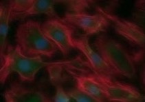
Cell transplantation is a major field in regenerative medicine and a promising alternative to whole organ transplantation. However, the process of cell engraftment is not yet fully understood and the hitherto achieved clinical outcome is limited. The aim of our study was to modify an aminosilan-coated nanoparticle for cell labeling and make it applicable for multimodal imaging using MRI, PET and fluorescent imaging. HIV-1 tat, linked FITC, and Gallium-68 were covalently bound to the particle and injected into Wistar rats. Animal-PET imaging was performed followed by MRI at 3.0T. Hepatic accumulation of the particles was proven by radionuclide distribution after 10 minutes in PET as well as in MRI over a 24 hour-period. Histological workup of the liver also revealed content of iron oxide particles in the reticuloendothelial system. Adjacent in vitro studies incubating hepatogenic HuH7 cells with the particles showed a rapid intracellular accumulation, clearly detectable by fluorescence microscopy and MRI. In conclusion our modified nanoparticle is stable under in vitro and in vivo conditions and is applicable for multimodal molecular imaging. Cellular labeling with this particle is possible and might help to get new insights into understanding the process of cell transplantation.
Read More
XXIII International Congress of TTS
Stacks Image 25549

The XXIII International Congress of The Transplantation Society, the leading international congress on transplantation biology and medicine, will be held in Vancouver, Canada from 15 - 19 August 2010. The translational program will draw together experts from around the world, and will encompass innovations in genomics and proteomics, molecular analyses of human diseases, innovations in biological and pharmacological immunosuppression, novel approaches to tolerance induction, technical advances in robotic surgery and imaging, advances in clinical practice and many other topics at the cutting edge of stem-cell, cellular and solid organ transplantation.
The XXIII Congress will be one of the largest and most exciting international gatherings of clinicians and scientists in the field of transplantation, drawing more than 4,000 delegates from all parts of the world. The international delegation will enjoy a stellar Scientific Program, commencing with a comprehensive and cutting-edge Postgraduate Education Program on Saturday 14th August and Sunday 15th August, and continuing with a panoply of outstanding plenary symposia, state of the art lectures, individual scientific presentations, and thematic symposia from Monday 16th August to Thursday 19th August inclusive.
The 2010 Congress will provide an outstanding opportunity for networking with colleagues and leading experts, and for sharing and discussing clinical advances, late-breaking discoveries and therapeutic advances in this field.
More information is available via www.transplantation2010.org. Furthermore, you may download the 2nd announcement here.
Important dates:
Abstract Submission Opens: October 2009
Online Registration Opens: November 2009
Abstract Submission Deadline: 5 March 2010
Author Notification of Acceptance: 7 May 2010
Early Registration Deadline: 4 June 2010
Late Breaking Abstract Deadline: 11 June 2010
Hotel Reservation Deadline: 25 June 2010
Regular Registration Deadline: 16 July 2010

Read More
Haluk Morgül defended thesis "magna cum laude"
Haluk Morgül successfully defended his medical doctoral thesis "magna cum laude".
After years of extremely fruitful research in the field of liver support, hepatocyte isolation and cell imaging via MRI he is (co-)author of 5 papers in peer reviewed journals (with more to come)!
Read More
cBAL111
Stacks Image 25570
Tanja Deurholt's paper on "Novel immortalized human fetal liver cell line, cBAL111, has the potential to differentiate into functional hepatocytes" is published in BMC Biotechnology. Co-authors are Niek P. van Til, Aniska A. Chhatta, Lysbeth ten Bloemendaal, Ruth Schwartlander, Catherine Payne, John N. Plevris, Igor M. Sauer, Robert A.F.M. Chamuleau, Ronald P.J. Oude Elferink, Jurgen Seppen, and Ruurdtje Hoekstra.
A clonal cell line that combines both stable hepatic function and proliferation capacity is desirable for in vitro applications that depend on hepatic function, such as pharmacological or toxicological assays and bioartificial liver systems. The article describes the generation and characterization of a clonal human cell line for in vitro hepatocyte applications.Cell clones derived from human fetal liver cells were immortalized by over-expression of telomerase reverse transcriptase. The resulting cell line, cBAL111, displayed hepatic functionality similar to the parental cells prior to immortalization, and did not grow in soft agar. Cell line cBAL111 expressed markers of immature hepatocytes, like glutathione S transferase and cytokeratin 19, as well as progenitor cell marker CD146 and was negative for lidocaine elimination. On the other hand, the cBAL111 cells produced urea, albumin and cytokeratin 18 and eliminated galactose. In contrast to hepatic cell lines NKNT-3 and HepG2, all hepatic functions were expressed in cBAL111, although there was considerable variation in their levels compared with primary mature hepatocytes. When transplanted in the spleen of immunodeficient mice, cBAL111 engrafted into the liver and partly differentiated into hepatocytes showing expression of human albumin and carbamoylphosphate synthetase without signs of cell fusion. This novel liver cell line has the potential to differentiate into mature hepatocytes to be used for in vitro hepatocyte applications.
Read More
World Conference on Regenerative Medicine 2009
Stacks Image 25575

The World Conference on Regenerative Medicine (WRM) will be held from October 29th to October 31st, 2009 in Leipzig. The Fraunhofer Institute for Cell Therapy and Immunology is once again organizer and host of this scientific event and the Translational Center for Regenerative Medicine (TRM) Leipzig has become co-organiser of the event. Main topics will be stem cells, tissue engineering, technology development, immunology, signaling, and regulatory affairs.
The final program is now available - more information via www.wcrm-leipzig.com .
Nathanael Raschzok will present our latest results concerning "Tracking of Transplanted Liver Cells in a Preclinical Large Animal Model", Thursday, October 29th, 2009, 04:00-05:30 pm, session: Imaging of Regeneration II, Hall 4.

Read More
ESAO 2009 - presentations
At the XXXVI Annual Meeting of the European Society for Artificial Organs ESAO 2009 in Compiègne/France the following projects have been presented by members of our group:

Tracking of transplanted liver cells via clinical 3.0 tesla MRI A. Zielinski, K. Steinz, N. Raschzok, N. Billecke, N. Kammer, M.H. Morgul, M. Adonopulou, S. Schmeisser, J. Pinkernelle, W. Ruedinger, U. Teichgraeber, I.M. Sauer
Evaluation of application sites for liver cell transplantation in a large animal model K. Steinz, A. Zielinski, N. Raschzok, N. Billecke, N.N. Kammer, M.H. Morgul, M. Adonopulou, L. Morawietz, W. Rüdinger, I.M. Sauer
Methylprednisolone and tacrolimus prevent hypothermia induced endothelial dysfunction - possible use in transplantation surgery K.R.L. Schmitt, A. Diestel, N. Billecke, F. Berger, I.M. Sauer
Read More
One Day of the Liver 2009
Stacks Image 25603
The traditional One Day on the Liver will be part of the XXXVI Annual Meeting of the European Society for Artificial Organs ESAO 2009 at the Université de Technologie de Compiègne in France. ODOL 2009 will take place September, 3rd focussing on "Cells, Hybrids, and Machines".
The following keynote lectures will be given:
Clinical Results of Liver Cell Therapy in Children with Urea Cycle Defect by Dr. Dr. W. Rüdinger, Weinheim, Germany,
Xenotransplantation. Where are we in 2009 ? by Dr. A. Billiau, Leuven, Belgium,
Problems and Opportunities: Perspectives of Bioartificial Liver Support by Dr. R. Hoekstra, Amsterdam, Netherlands,
New perspectives of artificial liver support by Prof. Dr. D. Falkenhagen, Krems, Austria.

We expect to present you all lectures as video streams after the meeting.
Read More
HAI 2009 - New aspects in liver support
Stacks Image 25613
The group was awarded with a research bonus (Forschungsprämie) by the Bundesministeriums für Bildung und Forschung (BMBF, Federal Ministry of Education and Research) supporting the development of micron sized particles for the detection of transplanted cells via MRI. The program is part of the High-Tech Strategy for Germany, launched by the Federal Government in August 2006 to encourage the development of new products and innovative services.
Read More
Nathanael Raschzok defended thesis "summa cum laude"
Stacks Image 25623
Nathanael Raschzok successfully defended his medical doctoral thesis "summa cum laude". After three years of extremely fruitful research and development in the field of hepatocyte transplantation, cell labeling, and MR imaging of transplanted cells he is first author of three papers in peer reviewed journals (with more to come...). He currently is finishing his in vivo MRI studies of MPIO labeled transplanted hepatocytes.
Transplantation of primary human hepatocytes is a promising approach in certain liver diseases. For visualisation of hepatocytes during and following cell application and the ability of a timely response to potential complications, a non-invasive modality for imaging of the transplanted cells has to be established. The aim of his studies was to label primary human hepatocytes with micron-sized iron oxide particles (MPIOs), enabling the detection of cells by clinical magnetic resonance imaging (MRI). The feasibility of preparing MPIO-labelled primary human hepatocytes detectable by clinical MR equipment was shown in vitro. MPIO-labelled cells could serve for basic research and quality control in the clinical setting of human hepatocyte transplantation.
He also investigated techniques for evaluation of the particle uptake via continuum source atomic absorption spectrometry (CSAAS). Porcine liver cells were labelled with MPIOs and the iron concentration of the cell samples was investigated by a CSAAS spectrometer equipped with a Perkin-Elmer THGA graphite furnace. CSAAS enabled rapid quantification of particle load from small quantities of cells without extensive preparation steps. CSAAS could be used for quality control in a clinical setting of cell transplantation.
Read More
Hypothermia induced endothelial dysfunction
Stacks Image 25633
Antje Diestel's manuscript entitled "Tacrolimus and methylprednisolone prevent hypothermia induced endothelial dysfunction" has been accepted for publication in the Journal of Heart and Lung Transplantation. Co-authors are Nils Billecke, Joerg Roessler, Boris Schmitt, Silke Troeller, Ruth Schwartlander, Felix Berger, Igor Maximilian Sauer and Katharina Rose Luise Schmitt.
Hypothermia is used to preserve organs for transplant and it is the oldest method to protect organs during complex pediatric cardiac surgery. Loss of tissue function and tissue edema are common complications in children undergoing cardiac surgery and heart transplantation. The present study was designed to examine the effects of methylprednisolone (MP) and Tacrolimus (TAC) on endothelial cell function and morphology after deep hypothermia and rewarming. Human umbilical vein endothelial cells (HUVECs) were pretreated with MP and/orTAC and incubated either within a specially designed bioreactor or in monolayers. They were then exposed to a dynamic cooling and rewarming protocol. Immunocytochemistry, time lapse video microscopy within the SlideReactor bioreactor system, cell permeability and adherence assays and western blot analysis were performed. Confluent endothelial cells exposed to hypothermia displayed elongated cell shapes with intercellular gap formation, increased endothelial cell-layer permeability and loss in adherence. Upon rewarming, however, endothelial cell integrity was restored. Opening and closing of intercellular gaps was dependent on ERK 1/2 activation and connexin 43 (Cx43) expression. The combined treatment with MP and TAC inhibited these hypothermia-induced changes. These results suggest that MP and TAC inhibit hypothermia induced endothelial gap formation via pERK 1/2 inhibition and connexin 43 stabilization. Application of combined drugs that affect multiple targets may therefore be considered as a possible new therapeutic strategy to prevent endothelial dysfunction after hypothermia and rewarming.
Read More
CSAAS and MPIO-labelled cells
Stacks Image 25643
As a result of the fruitful collaboration with the Institute for Analytical Sciences Berlin Nathanael Raschzok's paper on "Quantification of Cell Labelling with Micron-Sized Iron Oxide Particles Using Continuum Source Atomic Absorption Spectrometry" has been accepted by Tissue Engineering for publication. Co-authors are Nils Billecke, Nora N. Kammer, Mehmet H. Morgul, Michaela K. Adonopoulou, Igor M. Sauer, Stefan Florek, Helmut Becker-Ross, and Mao-Dong Huang.

Detection of cells after transplantation is necessary for quality control in regenerative medicine. Labelling with micron-sized iron oxide particles (MPIOs) enables non-invasive detection of single cells by magnetic resonance imaging. However, techniques for evaluation of the particle uptake are challenging. The aim of this study was to investigate continuum source atomic absorption spectrometry (CSAAS) for this purpose. Porcine liver cells were labelled with MPIOs and the iron concentration of the cell samples was investigated by a CSAAS spectrometer equipped with a Perkin-Elmer THGA graphite furnace. The weak iron line at 305.754 nm provides only about 1/600 sensitivity of the iron resonance line at 248.327 nm and was used for CSAAS measurements. Iron concentrations measured from labelled cells ranged from (5.8 ± 0.3) to (25.8 ± 0.9) pg Fe/cell, correlating to an uptake of (8.2 ± 0.5) to (25.7 ± 0.8) particles/cell. The results were verified by standardised morphometric evaluation. CSAAS enabled rapid quantification of particle load from small quantities of cells without extensive preparation steps. Thereby, CSAAS could be used for quality control in a clinical setting of cell transplantation.
Read More
Dr. med. Dominik Paul Modest
After years of research in the field of hepatocyte isolation and hypothermic long-term culture Dominik Paul Modest successfully defended his thesis "magna cum laude".
Read More
Dr. Igor M. Sauer: obtained postdoctoral lecture qualification (Habilitation) in Surgery
February 5th, 2009 at 4 p.m. (Hörsaal 3, Lehrgebäude Charité - Campus Virchow Klinikum) Dr. I.M. Sauer will give his inaugurative lecture entitled "Künstliche Organe: Von der Vision kybernetischer Organismen zur medizinischen Realität".
Read More
PhD Student Award in Regenerative Medicine
Stacks Image 25678
The Berlin-Brandenburg School for Regenerative Therapies (BSRT) offers young talented scientists in Berlin and Brandenburg:
Best Publication Award: The award will be granted for the most outstanding paper for progress in Regenerative Medicine accepted by a peer review journal in the last two years. The award comprises 1.500 Euro.
Best Presentation Award: The award will be granted for the best poster presented at an international conference with a peer review system within the last two years. The award comprises 500 Euro.
Info & Contact: BSRT, Charité Campus Virchow-Klinikum, Augustenburger Platz 1, 13353 Berlin, E-Mail: award@bsrt.de, Web: www.bsrt.de
Read More
Associated Investigator of BCRT
Stacks Image 25685
The Steering Committee of the Berlin-Brandenburg Center for Regenerative Therapies (BCRT) decided to involve Igor Sauer and his group as an Associated Investigator (AI).
The BCRT is a cooperative research institution of the Charite University Hospital in Berlin and Germany's largest research association, the Helmholtz Association. BCRT also receives generous financial support from the BMBF and the states of Berlin and Brandenburg, as well as from the Technology Foundations in Berlin and Brandenburg, the Future Fund Berlin and from various industry partners. More than 15 regional partners from science and industry are active members of the consortium at the BCRT.
Read More
Dr. med. Florian W.R. Vondran
The Steering Committee of the Berlin-Brandenburg Center for Regenerative Therapies (BCRT) decided to involve Igor Sauer and his group as an Associated Investigator (AI).
The BCRT is a cooperative research institution of the Charite University Hospital in Berlin and Germany's largest research association, the Helmholtz Association. BCRT also receives generous financial support from the BMBF and the states of Berlin and Brandenburg, as well as from the Technology Foundations in Berlin and Brandenburg, the Future Fund Berlin and from various industry partners. More than 15 regional partners from science and industry are active members of the consortium at the BCRT.
Read More
Imaging of human hepatocytes via MPIO and MRI
Stacks Image 25714
Nathanel Raschzok's and Haluk Morgül's manuscript entitled "Imaging of Primary Human Hepatocytes Using Micron-Sized Iron Oxide Particles and Clinical Magnetic Resonance Tomography" has been accepted for publication in the Journal of Cellular and Molecular Medicine (impact factor: 6,55). Authors are Nathanael Raschzok, Mehmet H. Morgul, Jens Pinkernelle, Florian W.R. Vondran, Nils Billecke, Nora N. Kammer, Gesine Pless, Michaela K. Adonopoulou, Christian Leist, Lars Stelter, Ulf Teichgraber, Ruth Schwartlander and Igor M. Sauer. Nathanael Raschzok and Mehmet Haluk Morgul contributed equally to this work. The contribution of Ruth Schwartländer has to be emphasised as well. Transplantation of primary human hepatocytes is a promising approach in certain liver diseases. For visualisation of hepatocytes during and following cell application and the ability of a timely response to potential complications, a non-invasive modality for imaging of the transplanted cells has to be established. The aim of this study was to label primary human hepatocytes with micron-sized iron oxide particles (MPIOs), enabling the detection of cells by clinical magnetic resonance imaging (MRI). Primary human hepatocytes isolated from 13 different donors were used for labelling experiments. Following dose finding studies, hepatocytes were incubated with 30 particles/cell for 4 hours in adhesion culture. Particle incorporation was investigated via light, fluorescence and electron microscopy and labelled cells were fixed and analysed in an agarose suspension by a 3.0 Tesla MR scanner. Hepatocytes were enzymatically resuspended and analysed during a five-day reculture period for viability, total protein, enzyme leakage (AST, LDH) and metabolic activity (urea, albumin). A mean uptake of 18 particles/cell could be observed, and primary human hepatocytes were clearly detectable by MR instrumentation. The particle load was not affected by resuspension and showed no alternations during the culture period. Compared to control groups, labelling and resuspension had no adverse effects on viability, enzyme leakage and metabolic activity of human hepatocytes. Conclusion: The feasibility of preparing MPIO-labelled primary human hepatocytes detectable by clinical MR equipment was shown in vitro. MPIO-labelled cells could serve for basic research and quality control in the clinical setting of human hepatocyte transplantation.
Read More
Tat-peptide modified MPIO
Stacks Image 25724
Haluk Morgül and Nathanael Raschzok published their first results on "Tracking of primary human hepatocytes with clinical MRI: Initial results with Tat-peptide modified superparamagnetic iron oxide particles." in the March issue of IJAO (Int J Artif Organs 2008, 31:252-257): The transplantation of primary human hepatocytes is a promising approach in the treatment of specific liver diseases. However, little is known about the fate of the cells following application. Magnetic resonance imaging (MRI) could enable real-time tracking and long-term detection of transplanted hepatocytes. The use of superparamagnetic iron oxide particles as cellular contrast agents should allow for the non-invasive detection of labelled cells on high-resolution magnetic resonance images. Experiments were performed on primary human hepatocytes to transfer the method of detecting labelled cells via clinical MRI into human hepatocyte transplantation. For labelling, Tat-peptide modified nano-sized superparamagnetic MagForce particles were used. Cells were investigated via a clinical MR scanner at 3.0 Tesla and the particle uptake within single hepatocytes was estimated using microscopic examinations. The labelled primary human hepatocytes were clearly detectable by MRI, proving the feasibility of this new concept. Therefore, this method is a useful tool to investigate the effects of human hepatocyte transplantation and to improve safety aspects of this method.
Read More
<<  Page 6 / 6 
M. Pflüger: Resect or not to resect: Unravelling the Biology of Neoplastic Pancreatic Cysts Using Genetic Studies
Stacks Image 19713

Michael Pflüger - currently research fellow at the Wood Lab, Sol Goldman Pancreatic Cancer Research Center, Department of Pathology, Johns Hopkins University School of Medicine - will present the translational work of the Wood Laboratory and the interdisciplinary management of pancreatic neoplasia at Johns Hopkins Hospital as part of the monthly interdisciplinary event series "The Pancreatic Cancer Precision Medicine Center of Excellence Program (PMCoE) Seminar Series".

"Resect or not to resect: Unravelling the Biology of Neoplastic Pancreatic Cysts Using Genetic Studies - A Translational Approach"
29.01.2024, 22:00/10 pm CET
Zoom link: https://jhjhm.zoom.us/j/94712957898?pwd=NXpHZ1NneFJtdTgvekx4ZVI1MHE3UT09

Archive


Categories

Year

Si-M | Topping-out Ceremony
Today, representatives of Charité – Universitätsmedizin Berlin and Technische Universität Berlin celebrated the topping-out ceremony for the research building "Der Simulierte Mensch" (Si-M, "The Simulated Human") together with political representatives. Guests included the Governing Mayor Franziska Giffey, Senator for Health and Science and Charité Supervisory Board Chair Ulrike Gote and Finance Senator Daniel Wesener.

We are very excited: this will be a great building with even greater content.

Stacks Image 15256
Si-M | Topping-out Ceremony
Today, representatives of Charité – Universitätsmedizin Berlin and Technische Universität Berlin celebrated the topping-out ceremony for the research building "Der Simulierte Mensch" (Si-M, "The Simulated Human") together with political representatives. Guests included the Governing Mayor Franziska Giffey, Senator for Health and Science and Charité Supervisory Board Chair Ulrike Gote and Finance Senator Daniel Wesener.

We are very excited: this will be a great building with even greater content.

Stacks Image 15266

This website or its third-party tools use cookies, which are necessary to its functioning and required to achieve the purpose illustrated in the Disclaimer. By closing this banner, scrolling this page, clicking a link or continuing to browse otherwise, you agree to the use of cookies.