Inaugural Lectures
We are pleased to announce that four members of staff have successfully completed their habilitation work in the last few months!

On
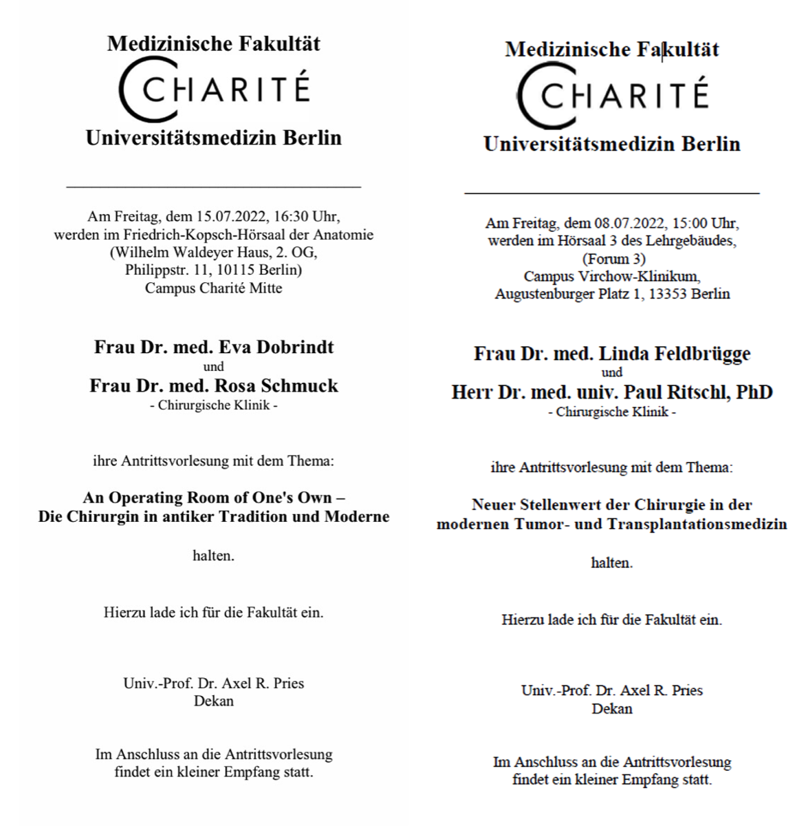
Friday, 08.07.2022 at 15:00 in lecture hall 3 of the teaching building (Forum 3, CVK), Dr. med. habil. Linda Feldbrügge and Dr. med. habil. Paul Ritschl will give their inaugural lectures entitled "New role of surgery in modern tumour and transplant medicine".

On
Friday, 15.07.2022 at 16:30 in the Friedrich Kopsch lecture theatre of the Anatomy Department at Campus Mitte Dr. med. habil. Eva Dobrindt and Dr. med. habil. Rosa Schmuck will present their inaugural lectures with the topic "An Operating Room of One's Own - The Surgeon in Ancient Tradition and Modernity".
This will be followed by a small reception in the park in front of the venue.
Stacks Image 28546
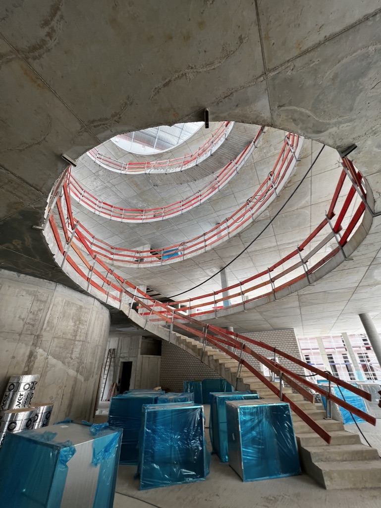
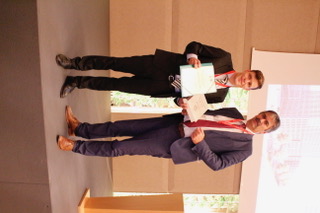
Si-M | Topping-out Ceremony
Today, representatives of Charité – Universitätsmedizin Berlin and Technische Universität Berlin celebrated the topping-out ceremony for the research building "Der Simulierte Mensch" (Si-M, "The Simulated Human") together with political representatives. Guests included the Governing Mayor Franziska Giffey, Senator for Health and Science and Charité Supervisory Board Chair Ulrike Gote and Finance Senator Daniel Wesener.

We are very excited: this will be a great building with even greater content.

Stacks Image 28556
Position for Student Assistant
Stacks Image 28607
Project: This project aims to analyse purinergic signal pathways, especially ecto-enzymes of the CD39 family and their role in tumor immunology. Tissue expression as well as the activity of the associated purinergic molecules (receptors, signaling molecules and degrading enzymes) will be examined in a patient cohort of different gastrointestinal tumors with and without (peritoneal) metastases.

Methods: Clinical tissue acquisition (perioperative of patients undergoing surgery), FACS, immunohistology, qPCR, cell culture.

Requirements: Medical student with clinical experience with patients (i.e. "Famulaturen") and drawing blood. Above average scientific interest and engagement. Teamwork and self sufficiency.

What we offer: Learning of scientific methods in an excellently equipped laboratory, teamwork. Mentoring in the clinical and lab. Publication of results and potential of a doctoral thesis.

Data: 10h/week. Project duration: 9 months. Begin: November 2021 (flexible). Salary according to TV Stud III for Berlin.

If you're the right person: please send all application documents, e.g. cover letter, curriculum vitae, certificates, attestations, etc. to the following address, quoting the reference number by e-mail to
Charité – Universitätsmedizin Berlin
Chirurgische Klinik, Exp. Chirurgie
z.Hd. Dr. Linda Feldbrügge
Augustenburger Platz 1
D-13353 Berlin
linda.feldbruegge@charite.de
Two new research grants by Berliner Krebsgesellschaft
Stacks Image 28617
The Berliner Krebsgesellschaft will fund two very interesting research projects by Dr. Linda Feldbrügge and Dr. Karl Hillebrandt in collaboration with Dr. Björn Papke.

Stacks Image 28624
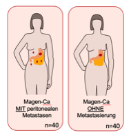
„Purinergic immune regulation in peritoneal metastases of gastric cancer via CD39 and ENTPD3 – target for a novel immune Checkpoint inhibition?“ – PI: Dr. Linda Feldbrügge

Peritoneal metastasis, especially derived from gastric cancer (GC), has a poor prognosis with a median survival of only months. Treatment is usually confined to palliative systemic chemotherapy, complemented individually by checkpoint inhibitors that block PD1-signaling. Innovative therapies combining surgery with local drug application such as hyperthermic intraperitoneal chemotherapy (HIPEC) or pressurized intraperitoneal aerosol chemotherapy (PIPAC) are still pending confirmation in clinical trials. Purinergic signaling, which involves ATP hydrolysis and generation of adenosine, regulated through CD39 (ENTPD1) and related enzymes, has been recognized as a critical immunoregulatory pathway in the tumor microenvironment (TME). The objective of the current project is to characterize the immune environment in the unique setting of peritoneal metastasis of gastric cancer with a focus on ectonucleotidases CD39 and ENTPD3 on T cells, macrophages and MDSC as well as mechanisms of ectonucleotidase-mediated immune regulation in tumor associated macrophages in vitro. As a high-volume center for surgical therapy of peritoneal malignancies and with years of experience in ectonucleotidase research, we aim to advance the understanding of peritoneal metastasis and contribute to improving treatment options for our patients.

Stacks Image 28631
"The influence of decellularised tumour matrix heterogeneity in relation to KRAS/MAPK inhibition of in vitro colorectal liver metastases." PI: Dr. Karl Hillebrandt and Dr. Björn Papke (Dept. of Pathology)

Colorectal cancer (CRC) is the second leading cause of cancer-related death worldwide, with approximately 900,000 annual deaths. 30-50% of patients develop colorectal liver metastases (CRLM) during their disease. More than 50% of these tumors have mutations in the KRAS oncogene, making them usually poorly treatable. Despite multimodal therapy concepts have improved the outcome of these patients, a large proportion of patients suffer a recurrence of their disease. For better therapeutic concepts, we need to better understand the tumor biology and metastatic mechanisms of these diseases. In vitro models, such as two-dimensional cell culture, are primarily used for this purpose. These models can only reflect the physiological complexity to a limited extent. Recently, it was shown that the use of organ-specific and tumor-specific extracellular matrix (ECM) has an impact on the behavior of human CRC cell lines. Culture of cell lines with decellularized matrix resulted in cells adopting a metastatic cell state and forming significantly more metastases in a mouse model than cells cultured on plastic or collagen. The goal of our project is to study the growth (with and without inhibition of the RAS/MAPK signaling pathway) of patient-derived tumor organoids growing on different decellularized metastatic matrices (dMM) and decellularized liver matrices (dLM). These studies of tumor matrix heterogeneity are essential to define which starting materials, for in vitro modeling of our three-dimensional tumor organoid culture, can be used to develop the most physiological, personalized dLM/dMM-based CRLM in vitro model possible. Based on these results, we plan to conduct small-scale therapy evaluations for personalized tumor therapy using our in vitro dLM/dMM-based CRLM in the near future.

Congratulations!
ADBoard | Therapeutic Assist and Decision Algorithms for Hepatobiliary Tumor Boards
Stacks Image 28645
The Gemeinsamer Bundesausschuss (Federal Joint Committee, G-BA) will fund a new collaborative project of the Charité's Dept. of Surgery and the Deutsches Forschungszentrum für Künstliche Intelligenz (German Research Center for Artificial Intelligence, DFKI), Speech and Language Technology.

The aim of the project Therapeutic Assist and Decision Algorithms for Hepatobiliary Tumor Boards (ADBoard) is the validation and evaluation of decision support systems based on linguistic and semantic methods of artificial intelligence (AI) for interdisciplinary tumour conferences in the care of tumour patients. Natural language processing (NLP) and machine learning (ML) will provide the technical basis for data extraction, data filtration and decision support for the automated generation of therapy recommendations. Interdisciplinary tumour board conferences are medical conferences, usually held on a weekly basis, which are required by the respective medical societies to determine a therapy or monitoring plan for patients with malignant diseases. Participants are representatives of the required medical disciplines who, taking into account the tumour characteristics and the general health of the patient, review the treatment options and make a therapy recommendation.

The Gemeinsamer Budesausschuss (Federal Joint Committee, G-BA) has the mandate to promote new forms of health care that go beyond the current standard provision of statutory health insurance, and health care research projects that are aimed at gaining knowledge to improve existing health care.

ADBoard is a collaboration of Priv.-Doz. Dr. Felix Krenzien, Priv.-Doz. Dr. Christian Benzing, Prof. Dr. Dominik Modest, Prof. Dr. Johann Pratschke (Charité – Universitätsmedizin Berlin) and Prof. Dr.-Ing. Sebastian Möller, Head of Research Department Speech and Language Technology, German Research Center for Artificial Intelligence.
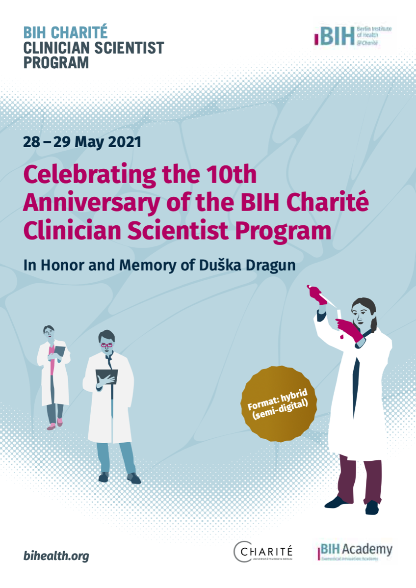
BIH Charité Clinician Scientist Symposium in Honor and Memory of Duška Dragun
Stacks Image 28654
28 May 2021 - 29 May 2021
BIH Charité Clinician Scientist Symposium in Honor and Memory of Duška Dragun

The symposium is composed of several components: First and foremost, it will commemorate Prof. Duška Dragun, the former Director of the BIH Biomedical Innovation Academy (BIA) and Director of the BIH Charité Clinician Scientist Program, who passed away in December 2020, and will be joined by stakeholders from academia and science policy. In addition, there will be scientific sessions, which will form tandems of program fellows and invited speaker. During a digital certificate ceremony on the evening of 28 May 2021, some 50 alumni will be bid farewell. The event language is English.

When
28 and 29 May 2021
10:00 - 6:30 pm

How
Online Event (semi-digital)

Registration
To receive the login link please register here.
Advanced Clinician Scientists
Stacks Image 28659
Priv.-Doz. Dr. Nathanael Raschzok and Priv.-Doz. Dr. Felix Krenzien successfully applied for the BIH Charité Advanced Clinician Scientist Pilot Programme (AdCSP) in a highly competitive process.

The BIH Charité AdCSP is designed as a career-phase-specific, sustainable funding programme that aims to closely interlink individual and institutional funding. The primary goal of the programme is to simultaneously incentivise the fellows and recognise the permissive academic culture of the respective clinics or institutes. Like the BIH Charité Clinician Scientist Programme (CSP) and the "Digital Clinician Scientist Programme" (DCSP), which has been additionally funded by the DFG since 2019, it is intended to be open to all clinical disciplines and to offer multiple networking opportunities for the funded fellows and participating clinics and institutes.

Congratulations!
Grant provided by the Berliner Krebsgesellschaft e.V.
Stacks Image 28664
Dr. med. M. Felsenstein receives a grant provided by the Berliner Krebsgesellschaft e.V. for his project "Deciphering the molecular determinants of pancreatic duct dysplasia by analysis of single-cell transcriptomics (RNAseq) in precursor lesions".

Besides great advances in the molecular and genetic understanding of pancreatic duct adenocarcinoma (PDAC), this tumor entity remains particularly aggressive with dismal prognosis. Recent single-cell sequencing studies underline the eminent urgency to understand tumor heterogeneity in the setting of PDAC. More detailed knowledge about the molecular mechanisms of pancreatic cancer evolution, carcinogenesis and heterogeneity could direct ideas for earlier detection and more effective targeted therapies, also preventing disease recurrence. Future therapeutic approaches in precision medicine will likely focus on the disease relevant sub-populations, specifically driving cancer progression, dissemination and exerting tumor escape mechanisms. In-depth transcriptomic data of single carcinoma environmental cells and respective cell clusters may help to discover novel biomarkers, which can be clinically instrumented for earlier detection and putatively increase the fraction of patients, amenable to curatively intended therapies. This study aims to analyze sorted single cells of macro-dissected precursor and cancerous lesions of the pancreas by single nuclei RNA sequencing (snRNAseq). In this feasibility study, we will include 10 patients, who will undergo resection of the pancreas due to “worrisome” or malignant lesions. We will perform in-depth transcriptomic analysis of pancreatic dysplasia in order to broaden our understanding of the molecular mechanisms of pancreatic carcinogenesis.

Congratulations!
Notch Signaling Pathway in Pancreatobiliary Tumors
Stacks Image 28684
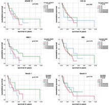
The Notch signaling pathway plays an important role both in the development of the ductal systems of the pancreas and the bile ducts as well as in cancer development and progression. The aim of this study was to examine the expression of central proteins of the Notch signaling pathway in pancreatobiliary tumors and its influence on patient survival.

Materials and Methods: We compared the receptors (Notch1, Notch4), activating splicing factors (ADAM17), and target genes (HES1) of the Notch pathway and progenitor cell markers with relevance for the Notch signaling pathway (CD44, MSI1) between pancreatic adenocarcinomas (PDAC, n = 14), intrahepatic cholangiocarcinoma (iCC, n = 24), and extrahepatic cholangiocarcinoma (eCC, n = 22) cholangiocarcinomas.

A significant overexpression of almost all studied components of the Notch signaling pathway can be found in the tumor tissue, however, without a significant influence on patient survival. Therefore, further studies are warranted to draw conclusions on Notch pathway's relevance for patient survival.

The paper "Notch Signaling Pathway in Pancreatobiliary Tumors" is available via Medicina, 2021;57(2):105. Authors are Francesca Borlak, Anja Reutzel-Selke, Anja Schirmeier, Julia Gogolok, Ellen von Hoerschelmann, Igor M Sauer, Johann Pratschke, Marcus Bahra, and Rosa B Schmuck.
Extended liver resection in mice: state of the art and pitfalls
Stacks Image 28689
"Extended liver resection in mice: state of the art and pitfalls – a systematic review" is available in ur J Med Res. 2021; 26(1):6.
Rodent models of liver resection have been used to investigate and evaluate the liver's complex physiology and pathology since 1931. First documented by Higgins and Anderson, such models were created to understand liver regeneration mechanisms to improve outcomes in patients undergoing extensive liver resection for liver cancer or other underlying liver diseases. A systematic search was conducted using Pubmed, gathering publications up to January 2019, which engaged with the mouse model of extended liver resection as a method itself. The results of this search were filtered according to their language, novelty, and relevancy.
Through the overview, laid out in the selected publications, this paper reviews the shift of the extended liver resection model from rat to the mouse, describes the state of the art in the experimental setting, and discusses the possible limitations and pitfalls. Clearly, the extended liver resection in mice is a reproducible, practical and easy to learn method.
Authors are Can Kamali, Kaan Kamali, Philipp Brunnbauer, Katrin Splith, Johann Pratschke, Moritz Schmelzle, and Felix Krenzien.
Duška Dragun
Stacks Image 28694
We have received the sad news that Professor Duška Dragun, Director of the Biomedical Innovation Academy (BIA) of the Berlin Institute of Health (BIH) and Head of the Charité BIH Clinician Scientist Program, succumbed to her long, severe, bravely endured illness on December 28, 2020 at the age of 51.
 
Her tireless efforts were devoted to her life's work: the Charité BIH Clinician Scientist Program. Ten years ago, she launched the first Clinician Scientist Program in Berlin and over the decade established and continuously expanded it as "best practice" for the German-speaking region. She has played a key role in developing and shaping the various programs for scientifically active physicians: from the Clinician Scientist Program, which enables aspiring specialists to spend up to 50 percent of their working time on research, to the Junior Clinician Scientist Program with 20 percent working time on research, which begins in the first year of specialist training, to the Advanced Clinician Scientist Program for specialists with postdoctoral qualifications. Two years ago, she successfully applied to the German Research Foundation (DFG) for the first and only Digital Clinician Scientist Program in Germany. This enables young physicians and scientists to conduct research and work in the field of digitalization in medicine and healthcare. Thus, within a few years, Duška Dragun made a significant contribution to building a new generation of young professionals for medicine – the impact of her programs will last for a long time, via promising individual careers as well as via the programmatic strengthening of a patient-oriented science.  
 
As a physician herself, Professor Duška Dragun has always been committed to research: As acting senior physician and deputy to the acting director of the Department of Nephrology and Intensive Care Medicine at the Charité Campus Virchow-Klinikum, as well as head of the nephrology research laboratory, she made highly regarded, internationally distinguished contributions to transplantation research with the goal of improving graft approach and long-term survival, preventing cardiovascular comorbidity, and thus improving the quality of life and life expectancy of transplanted patients.  She pursued her great goals with tremendous energy and passion, impressive perseverance and clear determination. She devoted her full attention to her employees, colleagues, and students, being equally attentive, understanding, and demanding.
 
The death of Duška Dragun is a great and painful loss. We will miss her greatly as director of the Charité BIH Clinician Scientist Program, as a physician, university professor and scientist. To us she was an inspiration, a mentor and an ever driving force.

Above all, however, we will greatly miss Duška as a friend.  
Karl Hillebrandt | Charité 3R Tandem project for early career researchers
Stacks Image 28699
Together with Dr. Björn Papke (Molecular tumour pathology), Dr. Karl Hillebrandt was able to acquire funding for a "Tandem project for early career researchers" from the Charité 3R. The project is entitled "A personalised therapy approach implementing individually matched matrix-based in vitro colorectal liver metastases to reduce metastatic mouse models".
Although modern multimodal therapy strategies have improved the clinical outcome of patients with colorectal liver metastases (CRLM), the overall prognosis is still poor. To further improve treatment options for patients, it is necessary to develop and test new targeted therapeutic approaches. To date, mouse models have often been used to study metastatic colorectal cancer. However, the rate of successful translation of animal models into clinical trials is less than 8%, highlighting the urgent need for alternative models to study the biology of metastatic cancer. This project aims to develop a novel personalised extracellular matrix-based in vitro model of human CRLM. This model will be validated against existing data from patient-derived organoids and xenografts (histology, single cell RNA sequencing and targeted gene sequencing). After internal comparison of our in vitro CRLM with the original CRLM, we will translate it into a personalised drug screening platform to test drug response from standard therapy to novel inhibitor combinations.
Felix Krenzien received Ferdinand-Sauerbruch Prize 2020
Stacks Image 28755
Priv.-Doz. Dr. Felix Krenzien received the Ferdinand-Sauerbruch Prize 2020 for his project and publication „The ILLS Laparoscopic Liver Surgery Fellow Skills Curriculum“ published in Annals of Surgery (online ahead of print).

Congratulations!

Laparoscopy is becoming the standard approach in liver surgery. As the degree of difficulty varies greatly from core skills to advanced procedures, strategies for teaching young surgeons need to be reconsidered. We here aimed to design a skills curriculum for LLR. Using the nominal group technique, 22 substeps of LLR were identified by 61 hepatobiliary surgeons. The raters were asked to rate (1) the difficulty of substeps and (2) the minimum number of times that the substep must be performed for mastery of the technique. According to the frequency of defined substeps, being estimated on the basis of high volume center experiences (n = 222 LLR; 1/2017-12/2018), the center's training capacity and defined goals for a 2-year fellowship were calculated.
Ten surgical substeps (45%) are routinely performed and can thus be taught sufficiently at centers carrying out ≥50 LLR in 2 years. As the mobilization of the right liver lobe and the dissection of the hepatic artery or portal vein is performed in only 27% and 28% of all LLR, respectively, sufficient training can only be provided at centers with ≥100 LLRs in 2 years. Mastery of complex parenchymal dissection (19%) and hilar lymphadenectomy (8%) can only be achieved in center performing ≥200 LLR in 2 years.
The authors suggest a stepwise approach for training of hepatobiliary fellows in LLR. Based on the estimated complexity of the substeps and the size of the center, not every substep can be learned within 2 years.

Authors are Felix Krenzien, Wenzel Schöning, Philipp Brunnbauer, Christian Benzing, Robert Öllinger, Matthias Biebl, Marcus Bahra, Nathanael Raschzok, Daniel Cherqui, David Geller, Ho-Seong Han, Go Wakabayashi, Moritz Schmelzle, Johann Pratschke, and the study group of the International Laparoscopic Liver Society (ILLS).
SiM | Der Simulierte Mensch
Stacks Image 28777
„Der Simulierte Mensch“ ("The Simulated Human", Si-M) is a new research building which is currently under construction and is expected to be finished in 2023. The building site is directly adjacent to the Charité Campus Virchow-Klinikum of the Charité - Universitätsmedizin in Berlin-Wedding and is also the birthplace of biotechnology at the TU Berlin.

The initiators of Si-M are Roland Lauster (Head of the Department of Medical Biotechnology at TU Berlin) and Andreas Thiel (Head of the research group Regenerative Immunology and Aging at Charité – Universitätsmedizin Berlin). They applied for the research building in 2018 (GG §91b) and successfully defended it before the German Science Council.

In the building, scientists from both institutions will work together to simulate the functions of human cells and tissues with new technologies of 3D cultivation, multi-organ chips or 3D bioprinting. In contrast to already existing collaborative projects, the building will be used to practice the joint development of models "side by side" in the same laboratory environment. In this way, both the development of organ models and technological developments can be adapted and optimized at the same time.

More information via https://www.si-m.org .

Stacks Image 28779
Magnetic resonance elastography quantification of decellularized liver tissue
Stacks Image 28816
"Magnetic resonance elastography quantification of the solid-to-fluid transition of liver tissue due to decellularization" was published in the latest issue of the Journal of the Mechanical Behavior of Biomedical Materials.

Maintenance of tissue extracellular matrix (ECM) and its biomechanical properties for tissue engineering is one of the substantial challenges in the field of decellularization and recellularization. Preservation of the organ-specific biomatrix is crucial for successful recellularization to support cell survival, proliferation, and functionality. However, understanding ECM properties with and without its inhabiting cells as well as the transition between the two states lacks appropriate test methods capable of quantifying bulk viscoelastic parameters in soft tissues.
We used compact magnetic resonance elastography (MRE) with 400, 500, and 600 Hz driving frequency to investigate rat liver specimens for quantification of viscoelastic property changes resulting from decellularization. Tissue structures in native and decellularized livers were characterized by collagen and elastin quantification, histological analysis, and scanning electron microscopy.
Decellularization did not affect the integrity of microanatomy and structural composition of liver ECM but was found to be associated with increases in the relative amounts of collagen by 83-fold (37.4 ± 17.5 vs. 0.5 ± 0.01 μg/mg, p = 0.0002) and elastin by approx. 3-fold (404.1 ± 139.6 vs. 151.0 ± 132.3 μg/mg, p = 0.0046). Decellularization reduced storage modulus by approx. 9-fold (from 4.9 ± 0.8 kPa to 0.5 ± 0.5 kPa, p < 0.0001) and loss modulus by approx. 7-fold (3.6 kPa to 0.5 kPa, p < 0.0001), indicating a marked loss of global tissue rigidity as well as a property shift from solid towards more fluid tissue behavior (p = 0.0097).
Our results suggest that the rigidity of liver tissue is largely determined by cellular components, which are replaced by fluid-filled spaces when cells are removed. This leads to an overall increase in tissue fluidity and a viscous drag within the relatively sparse remaining ECM. Compact MRE is an excellent tool for quantifying the mechanical properties of decellularized biological tissue and a promising candidate for useful applications in tissue engineering.

Authors are Hannah Everwien, Angela Ariza de Schellenberger, Nils Haep, Heiko Tzschätzsch, Johann Pratschke, Igor M. Sauer, Jürgen Braun, Karl H. Hillebrandt and Ingolf Sack.

J Mech Behav Biomed Mater. 2020 Apr;104:103640. doi: 10.1016/j.jmbbm.2020.103640. Epub 2020 Jan 14.
Characterization of pancreatic and biliary cancer stem cells in patient-derived tissue
Stacks Image 28821
Pancreatic ductal adenocarcinoma (PDAC) and extrahepatic cholangio-carcinoma (eCC) represent two cancer entities with devastating prognoses. Despite recent progress in research and treatment, therapy remains challenging. Cancer stem cells (CSCs) have been shown to play an important role in metastasis and chemoresistance. Therefore, CSCs may play a promising role as a potential therapeutic target.
A total of 31 patients (23 PDAC, 8 eCC) were included in the study. CSCs were analyzed in a single-cell suspension of tumor samples via fluorescence-activated cell scanning (FACS) with a functional Hoechst 33342 staining as well as a cell surface marker staining of the CSC-panel (CD24, CD44 and EpCAM) and markers to identify fibroblasts, leukocytes and components of the notch signaling pathway. Furthermore, the potential presence of CSCs among primary cancer-associated fibroblasts (CAFs) was assessed using the same FACS-panel.
We showed that CSCs are present in patient-derived dissociated tumor tissue. The functional and surface marker profile of CSC-detection did in fact correlate. The amount of CSCs was significantly correlated with tumor characteristics such as a higher UICC stadium and nodal invasion. CSCs were not restricted to the epithelial cell fraction in tumor tissues, which has been verified in independent analysis of primary cell cultures of CAFs.
Our study confirms the in vivo presence of CSCs in PDAC and eCC, stating a clinical significance thereof and thus their plausibility as therapeutic targets. In addition, stem-like cells also seem to constitute a part of the CAFs.

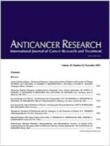
"Characterization of Pancreatic and Biliary Cancer Stem Cells in Patient-derived Tissue" was published in Anticancer Research. Authors are J. Gogolok, E. Seidel, A. Strönisch, A. Reutzel-Selke, I.M. Sauer, J. Pratschke, M. Bahra, and R.B. Schmuck.
New book: Decellularized Extracellular Matrix: Characterization, Fabrication and Applications
Stacks Image 28831
The extracellular matrix (ECM) supports cells and regulates various cellular functions in our body. The native ECM promises to provide an excellent scaffold for regenerative medicine. In order to use the ECM as a scaffold in medicine, its cellular fractions need to be removed while retaining its structural and compositional properties. This process is called decellularization, and the resulting product is known as the decellularized extracellular matrix (dECM).
The book Decellularized Extracellular Matrix: Characterization, Fabrication and Applications (Editors: Tetsuji Yamaoka, Takashi Hoshiba) focuses on the sources of dECM and its preparation, characterization techniques, fabrication, and applications in regenerative medicine and biological studies. Using this book, the reader will gain a good foundation in the field of ECM decellularization complemented with a broad overview of dECM characterization, ranging from structural observation and compositional assessment to immune responses against dECM and applications, ranging from microfabrication and 3D-printing to the application of tissue-derived dECM in vascular grafts and corneal tissue engineering etc. The book closes with a section dedicated to cultured cell dECM, a complementary technique of tissue-derived dECM preparation, for application in tissue engineering and regenerative medicine, addressing its use in stem cell differentiation and how it can help in the study of the tumor microenvironment as well as in clinical trials of peripheral nerve regeneration.

E. Keshi, I.M. Sauer and K.H. Hillebrandt contributed the chapter "Engineering an endocrine Neo-Pancreas".

The print version of this book (Royal Society of Chemistry, ISBN 978-1-78801-467-0) is planned for release on 11 December 2019.
TEBURU – our latest bioreactor system
Stacks Image 28858
Three‐dimensional tissue cultures are important models for the study of cell‐cell and cell‐matrix interactions, as well as, to investigate tissue repair and reconstruction pathways. Therefore, we designed a reproducible and easy to handle printable bioreactor system (Teburu), that is applicable for different approaches of pathway investigation and targeted tissue repair using human tissue slices as a three‐dimensional cell culture model. Here, we definitively describe Teburu as a controlled environment to reseed a 500‐µm thick decellularized human liver slice using human mesenchymal stroma cells. During a cultivation period of eight days, Teburu, as a semi‐open and low consumption system, was capable to maintain steady pH and oxygenation levels. Its combination with additional modules delivers an applicability for a wide range of tissue engineering approaches under optimal culture conditions.

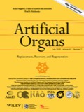
"Teburu—Open source 3D printable bioreactor for tissue slices as dynamic three‐dimensional cell culture models" was published in Artif Organs. 2019 Jun 18. doi: 10.1111/aor.13518. [Epub ahead of print]. Authors are A. Daneshgar, P. Tang, C. Remde, M. Lommel, S. Moosburner, U. Kertzscher, O. Klein, M. Weinhart, J. Pratschke, I.M. Sauer, and K.H. Hillebrandt.
Priv.-Doz. Dr. Felix Krenzien & Priv.-Doz. Dr. Christian Benzing
Stacks Image 28873
Today Dr. Felix Krenzien and Dr. Christian Benzing received they post-doctoral lecturing qualification (Habilitation) at Charité – Universitätsmedizin Berlin.
The title of Felix Krenzien's Habilitationsschrift is "Der differenzierte Einfluss der Seneszenz auf die Organtransplantation und Leberteilresektion", Christian Benzing focussed on the "Untersuchung der gesundheitsbezogenen Lebensqualität und der psychischen Gesundheit nach Lebertransplantation".

Congratulations !
Two new BIH Charité Junior Clinician Scientists
Stacks Image 28883
Dr. Karl Hillebrandt and Dr. Matthäus Felsenstein successfully applied for the BIH Charité Junior Clinician Scientist Program. Karl Hillebrandt will continue his work on human decellularized liver slices as 3D platform for in vitro models of cholangiocellular carcinoma. Matthäus Felsenstein focusses on derivation of normal pancreatic duct cells from human primary tissue and their stepwise genetic modification in vitro using CRISPR/Cas9 .

Congratulations!
Impact of Percoll purification on isolation of primary human hepatocytes
Stacks Image 28893
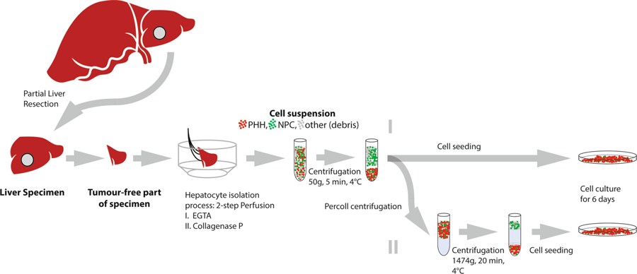
Scientific Reports published our paper "Impact of Percoll purification on isolation of primary human hepatocytes" (Sci Rep. 2019 Apr 25;9(1):6542). Authors are R. Horner, J.G.M.V. Gassner, M. Kluge, P. Tang, S. Lippert, K.H. Hillebrandt, S. Moosburner, A. Reutzel-Selke, J. Pratschke, I.M. Sauer, and N. Raschzok.

Research and therapeutic applications create a high demand for primary human hepatocytes. The limiting factor for their utilization is the availability of metabolically active hepatocytes in large quantities. Centrifugation through Percoll, which is commonly performed during hepatocyte isolation, has so far not been systematically evaluated in the scientific literature. 27 hepatocyte isolations were performed using a two-step perfusion technique on tissue obtained from partial liver resections. Cells were seeded with or without having undergone the centrifugation step through 25% Percoll. Cell yield, function, purity, viability and rate of bacterial contamination were assessed over a period of 6 days. Viable yield without Percoll purification was 42.4 × 106 (SEM ± 4.6 × 106) cells/g tissue. An average of 59% of cells were recovered after Percoll treatment. There were neither significant differences in the functional performance of cells, nor regarding presence of non-parenchymal liver cells. In five cases with initial viability of <80%, viability was significantly increased by Percoll purification (71.6 to 87.7%, p = 0.03). Considering our data and the massive cell loss due to Percoll purification, we suggest that this step can be omitted if the initial viability is high, whereas low viabilities can be improved by Percoll centrifugation.
Immunologic cellular characteristics of the tumour microenvironment of HCC
Stacks Image 28898
"Immunologic cellular characteristics of the tumour microenvironment of hepatocellular carcinoma drive patient outcomes" is available via World J Surg Oncol. 2019; 17(1):97

Anti-tumour immune competence has an impact in hepatocarcinogenesis and success of anti-cancer therapies. Tumour-infiltrating lymphocytes (TILs) and monocytes/macrophages (TAMs) are proposed to have significance in cancer. However, there is only limited data concerning their impact on patient outcome and survival in hepatocellular carcinoma (HCC).
Frequencies of CD68+, CD163+ M2-polarized TAMs and TILs were measured in de novo HCC tumours in non-cirrhosis (n = 58) using immunohistology and correlated to patients' clinicopathological characteristics and survival rates.
Patients with tumours marked by appearance of TILs and CD68+ TAMs showed an improved 1-, 3- and 5-year recurrence-free survival (all p ≤ 0.05). CD68+ TAMs were associated with reduced incidence of recurrent and multifocal disease. Conversely, CD163+ TAMs were associated with multifocal HCC and lymphangiosis carcinomatosa (all p ≤ 0.05).
TILs and CD68+ TAMs are associated with multiple tumour characteristics and patient survival in HCC. However, there is only scarce data about the biology underlying their mechanistic involvement in human tumour progression. Thus, experimental data on functional links might help develop novel immunologic checkpoint inhibitor targets for liver cancer.

Authors are G. Atanasov, K. Dino, K. Schierle, C. Dietel, G. Aust, J. Pratschke, D. Seehofer, M. Schmelzle, H.M. Hau.
Percoll purification after isolation of Primary Human Hepatocytes
Stacks Image 28934
The manuscript "Isolation of Primary Human Hepatocytes: Is Percoll Purification Really Necessary?" was accepted for publication in Scientific Reports.
Authors are Rosa Horner, Jospeh G.M.V. Gassner, Martin Kluge M, Peter Tang, Steffen Lippert, Karl H. Hillebrandt, Simon Moosburner, Anja Reutzel-Selke, Johann Pratschke, Igor M. Sauer and Nathanael Raschzok.

Research and therapeutic applications create a high demand for primary human hepatocytes. The limiting factor for their utilization is the availability of metabolically active hepatocytes in large quantities. Centrifugation through Percoll, which is commonly performed during hepatocyte isolation, has so far not been systematically evaluated in the scientific literature. 27 hepatocyte isolations were performed using a two-step perfusion technique on tissue obtained from partial liver resections. Cells were seeded with or without having undergone the centrifugation step through 25% Percoll. Cell yield, function, purity, viability and rate of bacterial contamination were assessed over a period of 6 days. Viable yield without Percoll purification was 42.4 x 10⁶ (SEM ± 4.6 x 10⁶) cells/g tissue. An average of 59% of cells were recovered after Percoll treatment. There were neither significant differences in the functional performance of cells, nor regarding presence of non-parenchymal liver cells. In five cases with initial viability of <80%, viability was significantly increased by Percoll purification (71.6 to 87.7%, p=0.03). Considering our data and the massive cell loss due to Percoll purification, we suggest that this step can be omitted if the initial viability is high, whereas low viabilities can be improved by Percoll centrifugation.
Read More
<<  Page 2 / 4  >>
Year
© 2025 Prof. Dr. Igor M. Sauer | Charité - Universitätsmedizin Berlin | Disclaimer

This website or its third-party tools use cookies, which are necessary to its functioning and required to achieve the purpose illustrated in the Disclaimer. By closing this banner, scrolling this page, clicking a link or continuing to browse otherwise, you agree to the use of cookies.