Proteomic profiling of colorectal liver metastases reveals histopathological response-specific molecular signatures of chemotherapy efficacy.
33 tissue samples from 31 patients were analyzed, including patients treated preoperatively with platinum-based chemotherapy, non-platinum-based chemotherapy, or targeted therapies, as well as untreated patients. Tumors were classified into major (MR), partial (PR), or no response (NR) groups using histopathological criteria. Proteomic profiling was performed using label-free mass spectrometry (LFQ-MS).
Analysis identified 607 proteins with differential expression across response types. Responsive CRLM were enriched in pathways related to immune infiltration, extracellular matrix organization, complement activation, and apolipoprotein processes, reflecting distinct stromal and immune patterns. Non-responsive tumors showed reduced expression of proteins involved in mitochondrial translation.
These findings indicate that CRLM have distinct proteomic phenotypes associated with histopathological response, largely independent of chemotherapy type, providing a foundation for biomarker discovery to predict and monitor chemotherapy efficacy.
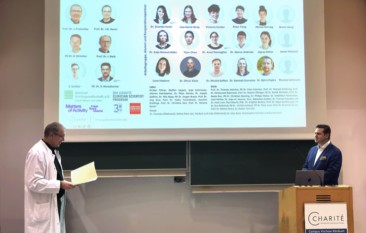
The paper is available in the March 2026 issue of Journal of Translational Medicine. Authors are A.K. Böhm, L.M. Skrip, D. Klein, F. Strobl, J.K. Wieland, A. Arnold, Y. Zhou, B. Papke, C. Sers, D.P. Modest, S. Moosburner, P.K. Haber, F. Krenzien, N. Raschzok, W. Schöning, D. Horst, T. Malinka, I. Sack, J. Pratschke, I.M. Sauer, and K.H. Hillebrandt.