News

Future Medicine 2017
Stacks Image 24555
What’s trending? What’s new in health science? To find out, please save the date for the second Future Medicine in Berlin on November 7, 2017. Tagesspiegel and Berlin Institute of Health, together with Charité - Universitätsmedizin Berlin and Max Delbrück Center for Molecular Medicine, will feature outstanding international scientists, great visions of the future of medicine, and an exceptional concentration of knowledge. The four sessions of Future Medicine 2017 will be: 

Digital Health and Big Data
Precision Medicine and Predictive Models
Cell and Gene Therapies
Stem Cells and Human Disease Modeling  

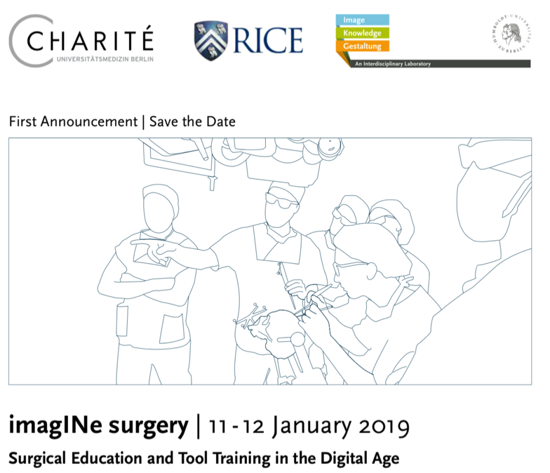
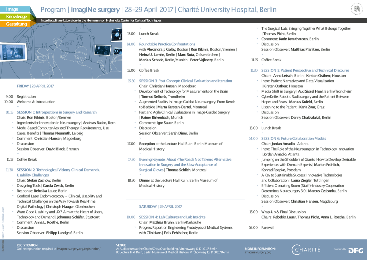
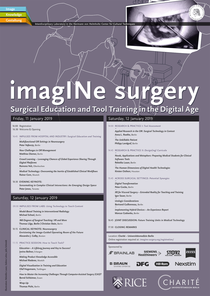
Simon Moosburner, a student of medicine interested in regenerative medicine and future technologies, will talk about “Virtual & Mixed Reality: The Next Milestone in Surgery?” at Future Medicine 2017.
Read More
Mixed Reality in Visceral Surgery
Stacks Image 24597
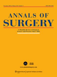
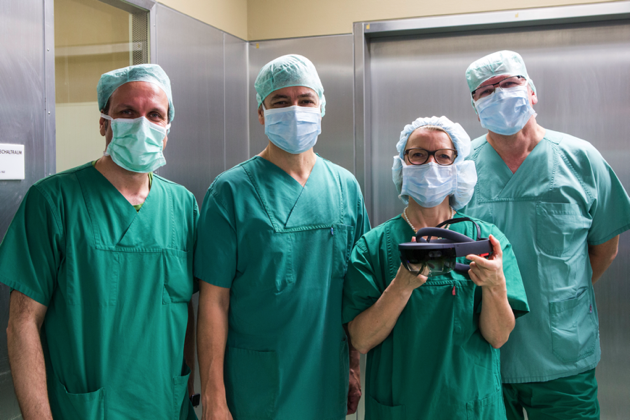
Annals of Surgery accepted our manuscript "Mixed Reality in visceral surgery - Development of a suitable workflow and evaluation of intraoperative use-cases" for publication. The paper evaluates the application of a mixed reality (MR) head-mounted display (HMD) for the visualization of anatomical structures in complex visceral-surgical interventions. A workflow was developed and technical feasibility was evaluated. 
Medical images are still not seamlessly integrated into surgical interventions and thus, remain separated from the surgical procedure. Surgeons need to cognitively relate two-dimensional sectional images to the three-dimensional (3D) during the actual intervention. MR applications simulate 3D images and reduce the offset between working space and visualization allowing for improved spatial-visual approximation of patient and image. The surgeon’s field of vision was superimposed with a 3D-model of the patient’s relevant liver structures displayed on a MR-HMD. This set-up was evaluated during open hepatic surgery. A suitable workflow for segmenting image masks and texture mapping of tumors, hepatic artery, portal vein and the hepatic veins was developed. The 3D model was positioned above the surgical site. Anatomical reassurance was possible simply by looking up. Positioning in the room was stable without drift and minimal jittering. Users reported satisfactory comfort wearing the device without significant impairment of movement. MR technology has high potential to improve the surgeon’s action and perception in open visceral surgery by displaying 3D anatomical models close to the surgical site. Superimposing anatomical structures directly onto the organs within the surgical site remains challenging since the abdominal organs undergo major deformations due to manipulation, respiratory motion and the interaction with the surgical instruments during the intervention. A further application scenario would be intraoperative ultrasound examination displaying the image directly next to the transducer. Displays and sensor-technologies as well as biomechanical modeling and object-recognition algorithms will facilitate the application of MR-HMD in surgery in the near future. Authors are I.M. Sauer, M. Queisner, P. Tang, S. Moosburner, O. Hoepfner, R. Horner, R. Lohmann and J. Pratschke.
Read More
Vascular anatomy of the juvenile Göttingen minipig
Stacks Image 24751
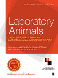
Lab Animal accepted our „Computed tomography-based survey of the vascular anatomy of the juvenile Göttingen minipig“ for publication.

Over the past 50 years, image-guided procedures have been established for a wide range of applications. The development and clinical translation of new treatment regimens necessitate the availability of suitable animal models. The juvenile Göttingen minipig presents a favourable profile as a model for human infants. However, no information can be found regarding the vascular system of juvenile minipigs in the literature. Such information is imperative for planning the accessibility of target structures by catheterization.

We present here a complete mapping of the arterial system of the juvenile minipig based on contrast-enhanced computed tomography. Four female animals weighing 6.13 ± 0.72 kg were used for the analyses. Imaging was performed under anaesthesia, and the measurement of the vascular structures was performed independently by four investigators. Our dataset forms a basis for future interventional studies in juvenile minipigs, and enables planning and refinement of future experiments according to the 3R (replacement, reduction and refinement) principles of animal research.


Authors are J. Siefert, K.H. Hillebrandt, M. Kluge, D. Geisel, P. Podrabsky, T. Denecke, M. Nösser, J. Gassner, A. Reutzel-Selke, B. Strücker, M.H. Morgul, S. Guel-Klein, J.K. Unger, A. Reske, J. Pratschke, I.M. Sauer, and N. Raschzok.
Read More
 Page 1 / 1 
Stacks Image 27655
Our manuscript "Depletion of donor dendritic cells ameliorates immunogenicity of both skin and hind limb transplants" has been accepted for publication in Frontiers in Immunology, section Alloimmunity and Transplantation. Authors are Muhammad Imtiaz Ashraf, Joerg Mengwasser, Anja Reutzel-Selke, Dietrich Polenz, Kirsten Führer, Steffen Lippert, Peter Tang, Edward Michaelis, Rusan Catar, Johann Pratschke, Christian Witzel, Igor M. Sauer, Stefan G. Tullius, and Barbara Kern.

Acute cellular rejection remains a significant obstacle affecting successful outcomes of organ transplantation including vascularized composite tissue allografts (VCA). Donor antigen presenting cells (APC), particularly dendritic cells (DC), orchestrate early alloimmune responses by activating recipient effector T cells. Employing a targeted approach, we investigated the impact of donor-derived conventional DC (cDC) and APC on the immunogenicity of skin and skin-containing VCA grafts, using mouse models of skin and hind limb transplantation.
By post-transplantation day 6, skin grafts demonstrated severe rejections, characterized by predominance of recipient CD4 T cells. In contrast, hind limb grafts showed moderate rejection, primarily infiltrated by CD8 T cells. While donor depletion of cDC and APC reduced frequencies, maturation, and activation of DC in all analysed tissues of skin transplant recipients, reduction in DC activities was only observed in the spleen of hind limb recipients. Donor cDC and APC depletion did not impact all lymphocyte compartments but significantly affected CD8 T cells and activated CD4 T in lymph nodes of skin recipients. Moreover, both donor APC and cDC depletion attenuated the Th17 immune response, evident by significantly reduced Th17 (CD4+IL-17+) cells in the spleen of skin recipients and reduced levels of IL-17E and lymphotoxin-α in the serum samples of both skin and hind limb recipients. In conclusion, our findings underscore the highly immunogenic nature of skin component in VCA. The depletion of donor APC and cDC mitigates the immunogenicity of skin grafts while exerting minimal impact on VCA.

Archive


Categories

Year

This website or its third-party tools use cookies, which are necessary to its functioning and required to achieve the purpose illustrated in the Disclaimer. By closing this banner, scrolling this page, clicking a link or continuing to browse otherwise, you agree to the use of cookies.