Prof. Dr. Moritz Queisner
Today Moritz Queisner received his appointment certificate for the professorship (W1) for Interdisciplinary Technology Transfer and Digitization in Surgery!
The professorship is associated with the DFG-funded Cluster of Excellence
»Matters of Activity«.

Congratulations!

Stacks Image 28459
On behalf of the Dean, Vice Dean Prof. Susanne Michl awards the certificate.
"Einstein Kickbox - Advanced Scientists" grant
Stacks Image 28468
Nils Haep successfully applied for the "Einstein Kickbox - Advanced Scientists" grant of the Einstein Center for Regenerative Therapies (ECRT). The purpose of the grant is to provide start-up funding for interesting experimental projects in regenerative medicine.  In his project, Nils is investigating the function of a cysteine-type endopeptidase and described mutations in the endopeptidase in hepatocytes using live-cell imaging and metabolomics. Through this preliminary work, he hopes to generate a hypothesis on the function of the endopeptidase and the underlying mechanism of the mutations. In the next step, he plans to develop a disease model for fatty liver from induced pluripotent stem cells.
Design Lab #13: Material Legacies
The exhibition »Design Lab #13: Material Legacies« at Kunstgewerbemuseum Berlin, opening on November 3rd, 2022, explores contingencies and ruptures between traditional crafts and the most recent developments at the crossroads of material research, design, engineering, and architecture. It brings together artifacts from the museum’s collection with work-in-progress installations by designers and researchers from the Cluster of Excellence »Matters of Activity. Image Space Material« in order to initiate a dialogue about the historical, contemporary, and future conditions under which materiality unfolds.

By engaging with a series of different materials and techniques the exhibition encompasses both the problematization of unsustainable pasts and presents as well as the imagination of speculative material futures. Taking materiality as a starting point, each of the exhibits will investigate its sociocultural, economic, and political context in order to disentangle the multiple interrelations that arise from and with materials. As such »Design Lab #13: Material Legacies« aims to challenge the passive understandings of materiality and associate with the widening discourse on relational knowledge practices in arts, design, humanities, and social science.

The exhibition will be running from 4 November 2022 to 26 February 2023. For the exhibition announcement on the website of the
Staatliche Museen zu Berlin – Preußischer Kulturbesitz.

Stacks Image 28478


Exhibition Opening

3 November 2022, 6 pm

The opening event will include an introduction to the exhibition by Dr. Claudia Banz, Curator of Design at the Kunstgewerbemuseum Berlin, and Prof. Dr. Claudia Mareis, co-director of the Cluster of Excellence »Matters of Activity. Image Space Material«. Moreover, exhibition curators Michaela Büsse and Emile De Visscher will provide background on the exhibition, its goals, and how the curatorial process was undertaken.
The exhibition opening is part of the Berlin Science Week 2022.
BMBF funds KIARA
With the programme "AI-based assistance systems for process-accompanying health applications", the Federal Ministry of Education and Research (BMBF) is funding innovative research and development work on interactive assistance systems that support processes in clinical health care using artificial intelligence methods.

Together with the partners Gebrüder Martin GmbH & Co. KG, Tuttlingen, HFC Human-Factors-Consult GmbH, Berlin and the Fraunhofer Institute for Telecommunications Heinrich-Hertz-Institut (HHI), Berlin, we successfully applied with the project "AI-based recording of work processes in the operating theatre for the automated compilation of the operating theatre report" (KIARA).


Stacks Image 28492


Operating theatre reports document all relevant information during surgical interventions. They serve to ensure therapeutic safety and accountability and as proof of performance. The preparation of the OR report is time-consuming and ties up valuable working time – time that is then not available for the treatment of patients.

In the KIARA project, we are working on a system that automatically drafts operating theatre reports. The KIARA system is intended to relieve medical staff: it documents operating theatre activities and creates a draft of the report, which then only needs to be checked, completed and approved. The system works via cameras integrated into operating theatre lamps. Their image data is then analysed with the help of artificial intelligence to recognise and record objects, people and all operating theatre activities. The ambitious system is to be developed and tested in a user-centred manner for procedures in the abdominal cavity and in oral and maxillofacial surgery.

KIARA is intended to continuously learn through human feedback and to simplify clinical processes for the benefit of medical staff by automating the creation of operating theatre reports. The system can also be applied to other operating theatre areas in the future.

The project has a financial volume of € 2.16 million.
The kick-off meeting took place on 16.09.2022 at the Charité.
„Si-M-Day“ | November 24th, 2022
Stacks Image 28502
Join us – at our online networking event.
We, the Si-M spokespersons and coordinators, are pleased to invite you to our first symposium „Si-M-Day“ on 24th November from 9 to 14 h – online.
It is dedicated to networking and initiation of projects between investigators of both partner institutions.
Click
here to register until November 18th (abstract submission deadline October 17th).
DFG Walter-Benjamin grant for the investigation of sex as a biological variable in alloimmunity
Stacks Image 28511
With a DFG Walter-Benjamin grant Dr. med. Friederike Martin will join the laboratory of Transplant Surgery Research at Harvard under the direction of Professor Stefan G. Tullius in Boston to investigate the role of biological sex for transplantation outcome.

Influences of donor and recipient sex on transplantation outcome have been described manifold, as well as an influence of sex hormones on the innate and adaptive immune response. So far, research, investigating the impact of sex hormones and different sex- and age-dependent sex-hormone levels on alloimmune response after solid organ transplantation is lacking. The aim of the project “Sex as a biological Variable in Alloimmunity” is, to delineate the impact of sex hormones and especially estrogens and age-dependent changes in estrogen-levels on alloimmune response after allogenic transplantation.  The project is based on the publication “Recipient sex and estradiol levels affect transplant outcomes in an age-specific fashion” published in the AJT in 2021 by the workgroup of Prof. Tullius.

Friederike, who already received the Sanofi Women in Transplantation fellowship grant for research in gender and sex in transplantation in 2021, will work as a Postdoc on this project in the Tullius Lab for an expected 2 years period, starting in January 2023.


Congratulations!
2022 TTS Mentee-Mentor-Award
Stacks Image 28517
Dr. Barbara Kern und Prof. Dr. Stefan G. Tullius received the 2022 Mentee-Mentor-Award of The Transplant Society during the 29th Conference in Buenos Aires.

In collaboration with National and International Societies, TTS acknowledges and recognizes the efforts of scientists who have advanced our understanding of transplantation science and fostered the development of young investigators.
The Mentee-Mentor Awards are designed to encourage dialogue and interactions between trainees and established investigators, and provide financial support for their joint participation in the Congress.

Congratulations!
Active Matter in Robotic-Assisted Surgery
Stacks Image 28529
Tuesday, 12.09.2022 | Cluster Retreat | Matters of Activity

2:30 – 2:45 pm Welcome & Intro
2:45 – 4:15 pm Panel 1
Rasa Weber Product Design (20 Minutes)
Felix Rasehorn Product Design (20 Minutes)
Binru Yang Engineering (20 Minutes)
Panel Discussion (30 Minutes)

4:15 – 4:45 pm Coffee Break
4:45 – 6:15 pm Panel 2
Jakub Rondomanski Mathematics (20 Minutes)
Babette Werner Art and Visual History (20 Minutes)
Anna Schäffner & Dominic Eger Domingos Product Design (20 Minutes)
Panel Discussion (30 Minutes)

6:15–7:30 pm Opening Exhibition und Aperitivo
Solid fraction determines stiffness and viscosity in decellularized pancreatic tissues
Stacks Image 28536
The article „Solid fraction determines stiffness and viscosity in decellularized pancreatic tissues“ in Biomaterials Advances is now available online.
There is free access to a PDF of the article here until August 20, 2022!

The role of extracellular matrix (ECM) composition and turnover in mechano-signaling and the metamorphic fate of cells seeded into decellularized tissue can be elucidated by recent developments in non-invasive imaging and biotechnological analysis methods. Because these methods allow accurate quantification of the composition and structural integrity of the ECM, they can be critical in establishing standardized decellularization protocols. This study proposes quantification of the solid fraction, the single-component fraction and the viscoelasticity of decellularized pancreatic tissues using compact multifrequency magnetic resonance elastography (MRE) to assess the efficiency and quality of decellularization protocols. MRE of native and decellularized pancreatic tissues showed that viscoelasticity parameters depend according to a power law on the solid fraction of the decellularized matrix. The parameters can thus be used as highly sensitive markers of the mechanical integrity of soft tissues. Compact MRE allows consistent and noninvasive quantification of the viscoelastic properties of decellularized tissue. Such a method is urgently needed for the standardized monitoring of decellularization processes, evaluation of mechanical ECM properties, and quantification of the integrity of solid structural elements remaining in the decellularized tissue matrix.

Authors are Joachim Snellings, Eriselda Keshi, Peter Tang, Assal Daneshgar, Esther C. Willma, Luna Haderer, Oliver Klein, Felix Krenzien, Thomas Malink, Patrick Asbach, Johann Pratschke, Igor M. Sauer, Jürgen Braun, Ingolf Sack, and Karl Hillebrandt.

Inaugural Lectures
We are pleased to announce that four members of staff have successfully completed their habilitation work in the last few months!

On
Friday, 08.07.2022 at 15:00 in lecture hall 3 of the teaching building (Forum 3, CVK), Dr. med. habil. Linda Feldbrügge and Dr. med. habil. Paul Ritschl will give their inaugural lectures entitled "New role of surgery in modern tumour and transplant medicine".

On
Friday, 15.07.2022 at 16:30 in the Friedrich Kopsch lecture theatre of the Anatomy Department at Campus Mitte Dr. med. habil. Eva Dobrindt and Dr. med. habil. Rosa Schmuck will present their inaugural lectures with the topic "An Operating Room of One's Own - The Surgeon in Ancient Tradition and Modernity".
This will be followed by a small reception in the park in front of the venue.
Stacks Image 28546
Si-M | Topping-out Ceremony
Today, representatives of Charité – Universitätsmedizin Berlin and Technische Universität Berlin celebrated the topping-out ceremony for the research building "Der Simulierte Mensch" (Si-M, "The Simulated Human") together with political representatives. Guests included the Governing Mayor Franziska Giffey, Senator for Health and Science and Charité Supervisory Board Chair Ulrike Gote and Finance Senator Daniel Wesener.

We are very excited: this will be a great building with even greater content.

Stacks Image 28556
VolumetricOR | Surgical Innovation
Stacks Image 28561
Our paper "VolumetricOR: A new Approach to Simulate Surgical Interventions in Virtual Reality for Training and Education" is available in the latest issue of Surgical Innovation.

Surgical training is primarily carried out through observation during assistance or on-site classes, by watching videos as well as by different formats of simulation. The simulation of physical presence in the operating theatre in virtual reality might complement these necessary experiences. A prerequisite is a new education concept for virtual classes that communicates the unique workflows and decision-making paths of surgical health professions (i.e. surgeons, anesthesiologists, and surgical assistants) in an authentic and immersive way. For this project, media scientists, designers and surgeons worked together to develop the foundations for new ways of conveying knowledge using virtual reality in surgery.
A technical workflow to record and present volumetric videos of surgical interventions in a photorealistic virtual operating room was developed. Situated in the virtual reality demonstrator called VolumetricOR, users can experience and navigate through surgical workflows as if they are physically present . The concept is compared with traditional video-based formats of digital simulation in surgical training.

VolumetricOR let trainees experience surgical action and workflows a) three-dimensionally, b) from any perspective and c) in real scale. This improves the linking of theoretical expertise and practical application of knowledge and shifts the learning experience from observation to participation.
Discussion: Volumetric training environments allow trainees to acquire procedural knowledge before going to the operating room and could improve the efficiency and quality of the learning and training process for professional staff by communicating techniques and workflows when the possibilities of training on-site are limited.

Authors are Moritz Queisner, Michael Pogorzhelskiy, Christopher Remde, Johann Pratschke, and Igor M. Sauer.
Tissue Engineering for the Diaphragm
"Tissue Engineering for the Diaphragm and its Various Therapeutic Possibilities – A Systematic Review" is available here in Advanced Therapeutics (open access).

Diaphragmatic impairments exhibit high morbidity as well as mortality while current treatment options remain unsatisfactory. Tissue engineering (TE) approaches have explored the generation of an optimal biocompatible scaffold for diaphragmatic repair through tissue decellularization or de novo construction, with or without the addition of cells. The authors conducted a systematic review on the current state of the art in diaphragmatic tissue engineering (DTE) and found 24 articles eligible for final synthesis. The included approaches studied decellularization-based graft generation and de novo bioscaffold construction. Three studies focused on in vitro host-scaffold interaction with synthesized, recellularized grafts and decellularized extracellular matrix scaffolds. Another three studies investigated evaluation tools for decellularization efficacy. Among all studies, recellularization is performed in both decellularization-based and de novo generated scaffolds. De novo constructed biocomposites as well as decellularized and recellularized scaffolds induced pro-regenerative remodeling and recovery of diaphragmatic function in all examined animal models. Potential therapeutic applications comprise substance defects requiring patch repair, such as congenital diaphragmatic hernia, and functional diseases demanding an entire organ transplant, like muscular dystrophies or dysfunction after prolonged artificial respiration.

Autors are Agnes K. Boehm, Karl H. Hillebrandt, Tomasz Dziodzio, Felix Krenzien, Jens Neudecker, Simone Spuler, Johann Pratschke, Igor M. Sauer, and Marco N. Andreas.
Stacks Image 28571
Detection of nicotinamide adenine dinucleotide (NAD) in cells and blood plasma
Stacks Image 28576
Priv,-Doz. Dr. med. Felix Krenzien and Dr. Jennifer Kirwan (Technologieplattform Metabolomik, Max-Delbrück-Centrum für Molekulare Medizin, Berlin) successfully applied for a grant within the Else Kröner Fresenius Stiftung funding line: Translational Research.

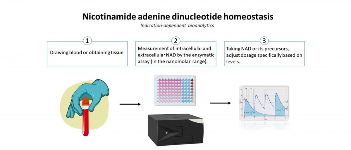
Recently, the molecule nicotinamide adenine dinucleotide (NAD) has attracted attention as it is involved in various important regulatory mechanisms, immune signaling, aging and regenerative processes. In this regard, it occupies key positions in many redox reactions of the body due to its role as a redox couple (NAD as an oxidized species and NADH as a reduced species). Consequently, NAD homeostasis (the maintenance of NAD in cells) is considered essential. The scientific consensus for many years was that the oxidized species resides exclusively in the intracellular milieu (iNAD). However, recent findings indicate that NAD also exists extracellularly (eNAD) and it is present in virtually all body fluids (from lymph to saliva to blood plasma). Based on these findings, precursors of NAD have recently been approved by the FDA and are commercially available. Measurement of eNAD in blood plasma is problematic due to its low concentration in the nanomolar range. However, quantifying eNAD plasma levels but also eNAD concentrations in cells is necessary to monitor the intake of NAD or its precursors and to adjust their dosage precisely.
The primary objective of this project is to validate, bioanalyze,and to document the assay for eNAD according to the ICH-M10 guidance document endorsed by the U. S. Food and Drug Administration (FDA). Adherence to the principles presented in this guideline should improve the quality and consistency of bioanalytical data, thereby supporting assay development and market approval. In addition, the assay will also be established for the measurement of intracellular NAD (iNAD), and validation of iNAD quantification will also be performed according to the guideline.

In the second part of the project, a clinical study will be conducted to determine whether the intake of nicotinamide riboside (precursor of NAD) leads to a change in eNAD and iNAD. Thus, the basis for an indication-dependent bioanalysis of the measurement of NAD will be developed to monitor the intake of NAD and its precursor or to adjust the dosage specifically on the basis of the quantification.
Stacks Image 28578

DFG Funds Extension of the Digital Clinician Scientist Program
Stacks Image 28587
Following the successful application for approval of the continuation of the BIH Charité Digital Clinician Scientist Program (DCSP) by the DFG, additional funding of around 1.3 million euros is now available over a period of two years: The DFG funding benefits physicians at Charité who have embarked on a scientific medical career path and, with their innovative research projects, are already playing a key role in shaping the digital transformation of healthcare during their residency training.

The Digital Clinician Scientist Program (DCSP) was jointly initiated by the German Research Foundation (DFG), the BIH, and the faculty of Charité - Universitätsmedizin Berlin in early 2019. The DCSP is an extension of the successful BIH Charité Clinician Scientist Program, which has set standards in the medical research landscape throughout Germany. The structured career path enables researching physicians to build the foundation for a successful career as a clinician scientist by providing protected time for research activities and non-clinical training during their residency. The DCSP is intended for clinically active physicians who are already actively shaping the digital transformation process of healthcare with their innovative research projects during their residency training. The main applicant of the continuation application is Prof. Dr. Igor M. Sauer, Director of the BIH Charité Digital Clinician Scientist Program, Deputy Clinic Director of the Department of Surgery, and Head of the Experimental Surgery at Charité. Since the start of the program in 2019, 24 physicians have benefited from funding, thus a broad spectrum of digital topics is already being addressed in various clinics at the Charité. The DFG originally funded the program for three years with more than three million euros. With the approved extension, the funding program now has a further 1.3 million euros available over a period of two years.
Development of human-based hydrogels as a substitute for mouse-derived Matrigel for cancer research
Stacks Image 28592
With a new funding line, Charité 3R wants to support the development of animal-free cell cultures at Charité.

For in vitro cancer research, mini-tumours are grown in a gel-like cultivation structure that serves the three-dimensional growth of the mini-tumours. This gel-like cultivation substance is obtained from mouse tumours, an unnatural cultivation environment for human mini-tumours. The aim of the project "Development of human-based hydrogels as a substitute for mouse-derived Matrigel for cancer research" by Björn Papke from the Institute of Pathology and Karl Hillebrandt is to produce a cultivation structure without animal additives. For this purpose, a cultivation structure, also gel-like, is to be produced from tissue obtained from patients during surgical procedures, which better corresponds to the natural environment of the human mini-tumours.


Congratulations!
 Page 1 / 1 
Year
© 2025 Prof. Dr. Igor M. Sauer | Charité - Universitätsmedizin Berlin | Disclaimer

This website or its third-party tools use cookies, which are necessary to its functioning and required to achieve the purpose illustrated in the Disclaimer. By closing this banner, scrolling this page, clicking a link or continuing to browse otherwise, you agree to the use of cookies.