Diffusion-weighted magnetic resonance imaging using a preclinical 1 T PET/MRI
Stacks Image 28977
"Diffusion-weighted magnetic resonance imaging using a preclinical 1 T PET/MRI in healthy and tumor-bearing rats" was published in EJNMMI Res. 2019 Feb 22;9(1):21. doi: 10.1186/s13550-019-0489-6.
Authors are J. Albrecht, D. Polenz, A.A. Kühl, J.M.M. Rogasch, A. Leder, I.M. Sauer, M. Babos, G. Mócsai, N. Beindorff, I.G. Steffen, W. Brenner, and E.J. Koziolek.

Hybrid positron emission tomography and magnetic resonance imaging (PET/MRI) scanners are increasingly used for both clinical and preclinical imaging. Especially functional MRI sequences such as diffusion-weighted imaging (DWI) are of great interest as they provide information on a molecular level, thus, can be used as surrogate biomarkers. Due to technical restrictions, MR sequences need to be adapted for each system to perform reliable imaging. There is, to our knowledge, no suitable DWI protocol for 1 Tesla PET/MRI scanners.
We established a respiratory-gated DWI protocol for a preclinical 1 T PET/MRI scanner allowing to monitor growth-related changes in ADC values of orthotopic HCC liver tumors. By monitoring the changes in tumor ADCs over time, different cellular stages were described. However, each study needs to adapt the protocol further according to their question to generate best possible results.
Read More
Charité Digital Clinician Scientist Pilot Program (D-CSP)
The Deutsche Forschungsgemeinschaft (DFG) will fund the Charité Digital Clinician Scientist Pilot Program (D-CSP). The ideas is to improve and safeguard the current BIH Charité Clinician Scientist Program by building an additional structure for a novel “digital science” driven career track to prepare academic clinicians for the challenges of the emerging technological transformation of medicine.
Stacks Image 28988
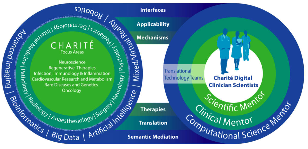
Designated Spokesperson is Prof. Dr. Duska Dragun. Co-applicants are the NeuroCure Cluster of Excellence, Department of Experimental Neurology, Department of Pediatric Oncology and Hematology, Department of Radiology and Pediatric Radiology, Department of Surgery, Berlin Institute for Medical Systems Biology (BIMSB), Institute of Medical Biometrics and Clinical Epidemiology, Department of Neurology and Experimental Neurology, and the Department of Anesthesiology and Intensive Care Medicine.

With the changing dynamics in biomedical research having fully entered into the digital era, it is becoming increasingly clear after seven years of experience that we need more dedicated efforts to create opportunities by establishing stronger interfaces with physics, mathematics, systems biology, and computational sciences for future generations of Clinician Scientists. The newly proposed research and educational structure for integrating these new areas of expertise into the established CSP should act as a “central processing unit” to facilitate biomedical knowledge derived from a variety of clinical disciplines supported by leading technology experts to address the specific challenges of data-driven medicine in the future.

  • Precision medicine in cancer and beyond,
  • Systems biology,
  • Big data science and decision support systems,
  • Quantitative imaging,
  • Computational neuroscience and brain simulation, and
  • Augmented, mixed and virtual reality in surgery
are exemplary research topics highlight how applicants will interact with Digital Clinician Scientists to develop their skills in giving prognoses, optimizing delivery of care, and personalizing patient management and therapeutic choices.
Read More
Mathilde Feist and Paul Ritschl: Clinician Scientists
Stacks Image 29037
Dr. Mathilde Feist and Dr. Paul Ritschl successfully applied for the BIH Charité Clinician Scientist Program. Mathilde Feist will work on "Cytokine-armed oncolytic vaccinia virus for pancreatic cancer therapy". Mentors are Prof. Bahra, Prof. Sauer and Prof. Beling.
Paul Ritschl focusses on "The Impact of Donor Derived Microparticles Following Solid Organ Transplantation". Mentors are Priv.-Doz. Dr. Schmelzle and Priv.-Doz Dr. Öllinger.
Read More
Nanomolar sensing of NAD
Stacks Image 29052
"The nanomolar sensing of nicotinamide adenine dinucleotide in human plasma using a cycling assay in albumin modified simulated body fluids." was published in Nature Scientific Reports.
Authors are P. Brunnbauer, A. Leder, C. Kamali, K. Kamali, E. Keshi, K. Splith, S. Wabitsch, P. Haber, G. Atanasov, L. Feldbrügge, I.M. Sauer, J. Pratschke, M. Schmelzle, and F. Krenzien.

Nicotinamide adenine dinucleotide (NAD), a prominent member of the pyridine nucleotide family, plays a pivotal role in cell-oxidation protection, DNA repair, cell signalling and central metabolic pathways, such as beta oxidation, glycolysis and the citric acid cycle. In particular, extracellular NAD+ has recently been demonstrated to moderate pathogenesis of multiple systemic diseases as well as aging. Herein we present an assaying method, that serves to quantify extracellular NAD+ in human heparinised plasma and exhibits a sensitivity ranging from the low micromolar into the low nanomolar domain. The assay achieves the quantification of extracellular NAD+ by means of a two-step enzymatic cycling reaction, based on alcohol dehydrogenase. An albumin modified revised simulated body fluid was employed as standard matrix in order to optimise enzymatic activity and enhance the linear behaviour and sensitivity of the method. In addition, we evaluated assay linearity, reproducibility and confirmed long-term storage stability of extracellular NAD+ in frozen human heparinised plasma. In summary, our findings pose a novel standardised method suitable for high throughput screenings of extracellular NAD+ levels in human heparinised plasma, paving the way for new clinical discovery studies.
Read More
Tumor-stromal cross-talk in PDAC
Stacks Image 29074
Hepatobiliary Pancreat Dis Int accepted the manuscript "Tumor-stromal cross-talk modulating the therapeutic response in pancreatic cancer" for publication. Authors are C.C.M. Neumann, E. von Hörschelmann, A. Reutzel-Selke, E. Seidel, I.M. Sauer, J. Pratschke, M. Bahra, and R.B.Schmuck.

Pancreatic ductal adenocarcinoma (PDAC) is a highly malignant solid tumor with a dismal prognosis. The stroma component makes up to 90% of the tumor mass and is thought to be one of the main reasons for the tumor's high chemoresistance. Cancer associated fibroblasts (CAFs) have previously been identified to be the key stromal players. This is the first time we provide detailed in vitro experiments investigating tumor-stromal interactions when exposed to three well-known chemotherapeutic agents.
Monocultures, indirect and direct co-cultures of two PDAC cell lines (AsPC and Panc-1) and six primary patients derived CAFs were treated with gemcitabine, nab-paclitaxel and the γ-secretase-inhibitor (GSI) DAPT. The cell viability of each component was measured with XTT. Finally, IL-6 concentrations of the supernatants were analyzed.
On the contrary to PDAC cell lines, CAF monocultures hardly responded to any treatment which suggested that stroma (CAFs) itself is more resistant to standard chemo-treatments than the epithelial cancer cells. Moreover, only a weak chemotherapeutic response was observed in direct co-cultures of cancer cells with CAFs. A change in the morphology of direct co-cultures was accompanied with the chemoresistance. CAFs were observed to build cage-like structures around agglomerates of tumor cells. High levels of IL-6 were also associated with a reduced response to therapy. Indirect co-cultures make the tumor-stromal interaction more complex.
Read More
Prof. Marcus Bahra Charité W2 professor
Stacks Image 29081
Prof. Dr. Marcus Bahra was appointed as a W2 professor for Pancreato-Biliary Surgery at the Charité – Universitätsmedizin Berlin. Working with other specialists, his team provides treatment for patients with malignancies and diseases in the pancreas and bile duct and conducts research in this field.
Since 2014 he held an extraordinary professorship for Surgery at the Charité.
Read More
Isolation of primary human hepatocytes & LiMax-test
Stacks Image 29111
Tissue Engineering (Part C: Methods) accepted our paper entitled "The predictive value of the LiMAx-test for the isolation of primary human hepatocytes".
Authors are R.D. Major, M. Kluge, M. Jara, M. Nösser, R. Horner, J. Gassner, B. Struecker, P. Tang, S. Lippert, A. Reutzel-Selke, D. Geisel, T. Denecke, M. Stockmann, J. Pratschke, I.M. Sauer, and N. Raschzok.

The need for primary human hepatocytes is constantly growing, for basic research as well as for therapeutic applications. However, the isolation outcome strongly depends on the quality of liver tissue, and we are still lacking a preoperative test that allows the prediction of the hepatocyte isolation outcome. Here we evaluated the “maximal liver function capacity test” (LiMAx) as predictive test for the quantitative and qualitative outcome of hepatocyte isolation. This test is already used in clinical routine to measure preoperative and to predict postoperative liver function.
The patient’s preoperative mean LiMAx was obtained from the patient records and preoperative CT and MRI images were used to calculate the whole liver volume in order to adjust the mean LiMAx. The outcome parameters of the hepatocyte isolation procedures were analyzed in correlation with the adjusted mean LiMAx.
Primary human hepatocytes were isolated from partial hepatectomies (n=64).
From these 64 hepatectomies we included 48 to our study and correlated their isolation outcome parameters with volume corrected LiMAx values. From a total of 11 hepatocyte isolation procedures, metabolic parameters (albumin, urea and aspartate aminotransferase) were assessed during the hepatocyte cultivation period of 5 days. The volume adjusted mean LiMAx showed a significant positive correlation with the total cell yield (p= 0.049;r= 0.242;n= 48). The correlations of volume adjusted LiMAx values with viable cell yield and cell viability did not reach statistical significance. A sub-group analysis of isolations from patients with colorectal metastasis revealed a significant correlation between volume adjusted mean LiMAx and total cell yield (p= 0.012;r= 0.488;n= 21) and viable cell yield (p=0.034;r=0.405;n=21). Whereas a sub-group analysis of isolations of patients with carcinoma of the biliary tree showed significant correlations of volume adjusted mean LiMAx with cell viability (r= 0.387;p= 0.046;n=20) and lacked significant correlations with total cell yield (r= - 0.060;p= 0.401;n=20) and viable cell yield (r= 0.012;p= 0.480;n=20). The volume-adjusted mean LiMAx did not show a significant correlation with any of the metabolic parameters. In conclusion, the LiMAx-test might be a useful tool to predict the quantitative outcome of hepatocyte isolation, as long as underlying liver disease is taken into consideration.
Read More
ECRT Kickbox - Junior Scientist Grant
Stacks Image 29247
Karl Hillebrandt receives one of the 2017 Einstein Center for Regenerative Therapies (ECRT) Kickbox – Junior Scientist Grant. The project is entitled "Fighting liver cirrhosis? Establishment and analysis of decellularized human cirrhotic liver slices as a 3-dimensional model to study cell matrix interactions".

Liver cirrhosis is one of the main indications for liver transplantation. Due to the organ shortage, this therapy option is limited to the minority of patients suffering from cirrhosis. Therefore, there is a need of alternative treatment options.The aim of our project is to establish a decellularization protocol for human cirrhotic livers slices, which preserves the natural extracellular matrix (ECM) of cirrhotic livers. These decellularized liver slices will serve as a 3 dimensional model to study cell matrix interactions. If we are able to establish a protocol which will preserve the ECM, we will conduct in vitro recellularization experiments to study how the cirrhotic ECM will change the genotype and phenotype of different cell types. With this knowledge we aim to modify specific cell types in vivo or vitro for example prior to cell transplantation. Our ambition is to steer the cell matrix interaction via these modified cells after their transplantation and thereby halt or even reverse the progress of liver cirrhosis. This approach may offer an alternative treatment option in the future.

Team : Karl Hillebrandt, Oliver Klein, Ben Strücker, Igor Sauer  
Read More
Vascular anatomy of the juvenile Göttingen minipig
Stacks Image 29324
Lab Animal accepted our „Computed tomography-based survey of the vascular anatomy of the juvenile Göttingen minipig“ for publication.

Over the past 50 years, image-guided procedures have been established for a wide range of applications. The development and clinical translation of new treatment regimens necessitate the availability of suitable animal models. The juvenile Göttingen minipig presents a favourable profile as a model for human infants. However, no information can be found regarding the vascular system of juvenile minipigs in the literature. Such information is imperative for planning the accessibility of target structures by catheterization.

We present here a complete mapping of the arterial system of the juvenile minipig based on contrast-enhanced computed tomography. Four female animals weighing 6.13 ± 0.72 kg were used for the analyses. Imaging was performed under anaesthesia, and the measurement of the vascular structures was performed independently by four investigators. Our dataset forms a basis for future interventional studies in juvenile minipigs, and enables planning and refinement of future experiments according to the 3R (replacement, reduction and refinement) principles of animal research.


Authors are J. Siefert, K.H. Hillebrandt, M. Kluge, D. Geisel, P. Podrabsky, T. Denecke, M. Nösser, J. Gassner, A. Reutzel-Selke, B. Strücker, M.H. Morgul, S. Guel-Klein, J.K. Unger, A. Reske, J. Pratschke, I.M. Sauer, and N. Raschzok.
Read More
HCC: miRNA profiles of the tumor-surrounding tissue
Stacks Image 29351
The presence of hepatocellular carcinoma (HCC) is a significant complication of cirrhosis because it changes the prognosis and the treatment of the patients. By now, contrast-enhanced CT and MR scans are the most reliable tools for the diagnosis of HCC; however, in some cases, a biopsy of the tumor is necessary for the final diagnosis. The aim of the study was to develop a diagnostic tool using the microRNA (miRNA) profiles of the tissue surrounding the HCC tumor combined with clinical parameters in statistical models. At a transplantation setting, 32 patients with HCC and cirrhosis (B) were compared to 22 patients suffering from cirrhosis only (A). The diagnosis and exclusion of HCC was confirmed following the histopathological examination of the explanted liver.

The HCC patients were significantly older than the patients with cirrhosis only (B: 60.6 and A: 49.9, p<0.001) and showed higher levels of ALT (A: 0.76μkat/l, B: 1.02μkat/, p=0.006) and AFP (A: 5.8ng/ml, B: 70.3ng/ml, p<0.001), whereas the bilirubin levels were higher in the cirrhosis only group (p=0.002). Using age (cut-off 50.23years) and AFP (cut-off 4.2ng/ml) thresholds, the levels of expression of miR-1285-3p and miR-943 differentiated between the patients with HCC and cirrhosis from those with cirrhosis only with an accuracy of 96.3%. This is the first report about the use of stepwise penalized logistic regression and decision tree analyses of miRNA expressions in the tumor-surrounding tissue combined with clinical parameters for the diagnosis of HCC.

Authors are Mehmet Haluk Morgul, Sergej Klunk, Zografia Anastasiadou, Ulrich Gauger, Corinna Dietel, Anja Reutzel-Selke, Philipp Felgendref, Hans-Michael Hau, Hans-Michael Tautenhahn, Rosa Bianca Schmuck, Nathanael Raschzok, Igor Maximillian Sauer, and Michael Bartels. Exp Mol Pathol. 2016 Aug 20;101(2):165-171. doi: 10.1016/j.yexmp.2016.07.014. [Epub ahead of print]
Read More
Hepatocyte isolation after laparoscopic liver resection
Stacks Image 29369
Tissue Engineering, Part C: Methods accepted our paper entitled "Hepatocyte isolation after laparoscopic liver resection" for publication. Authors are Horner R*, Kluge M*, Gassner J, Nösser M, Major RD, Reutzel-Selke A, Leder AK, Struecker B, Morgul MH, Pratschke J, Sauer IM, Raschzok N (*contributed equally).

Liver tissue obtained from partial hepatectomy is a common source for isolation of primary human hepatocytes. Until now, liver resections were most commonly performed by conventional open surgery. Although the laparoscopic approach is currently emerging in liver surgery, data on the outcome of hepatocyte isolation from laparoscopically resected liver tissue is not available. A total of 22 hepatocyte isolations were performed using the two-step collagenase perfusion technique from October 2015 until March 2016. Liver tissue was obtained from n=15 open liver resections (OLR) and n=7 laparoscopic liver resections (LLR). Isolation parameters (cell yield, viability, percoll survival) were assessed and hepatocyte function (plating efficiency, urea, albumin, and aspartate aminotransferase) was measured over a culture period of 6 days (OLR: n=13; LLR: n=3). Total cell yield (OLR: 36.81 ± 6.77 x10⁶ cells/g vs. LLR 16.84 ± 10.66 x10⁶ cells/g, p=0.0318) as well as viable yield (OLR 31.70 ± 6.05 x10⁶ cells/g vs. LLL 14.70 ± 9.89 x10⁶ cells/g, p=0.0260) were significantly higher in the OLR group. Subgroup analysis revealed that the worse outcome of isolation of laparoscopically resected liver tissue was associated with right-lateral laparoscopic liver resections, while hepatocyte isolation from left-lateral laparoscopic liver resections was as effective as from open surgery. Hepatocyte function did not differ between hepatocytes from openly resected versus left-lateral laparoscopically resected liver tissue. We here present the first data on hepatocyte isolation from laparoscopic liver surgery. While the overall outcome is worse compared to open surgery, our data suggest that liver tissue from laparoscopic resection of the left lobe is an excellent source for primary human hepatocytes.
Read More
Human hepatocyte isolation – new paper
Stacks Image 29484
Tissue Engineering, Part C: Methods accepted our paper „Human hepatocyte isolation: Does portal vein embolization affect the outcome?“ Authors are Martin Kluge, Anja Reutzel-Selke, Hendrik Napierala, Karl H. Hillebrandt, Rebeka D. Major, Benjamin Struecker, Annekatrin Leder, Jeffrey Siefert, Peter Tang, Steffen Lippert, Daniel Seehofer, Johann Pratschke, Igor M. Sauer und Nathanael Raschzok.

Primary human hepatocytes are widely used for basic research, pharmaceutical testing, and therapeutic concepts in regenerative medicine. Human hepatocytes can be isolated from resected liver tissue. Preoperative portal vein embolization (PVE) is increasingly used to decrease the risk of delayed postoperative liver regeneration by induction of selective hypertrophy of the future remnant liver tissue. The aim of this study was to investigate the effect of PVE on the outcome of hepatocyte isolation. Primary human hepatocytes were isolated from liver tissue obtained from partial hepatectomies (n=190) using the two-step collagenase perfusion technique followed by Percoll purification. Of these hepatectomies, 27 isolations (14.2%) were performed using liver tissue obtained from patients undergoing PVE prior to surgery. All isolations were characterized using parameters that had been described in the literature as relevant for the outcome of hepatocyte isolation. The PVE and non-PVE groups were similar in regard to donor parameters (sex, age, indication for surgery), isolation parameters (liver weight, cold ischemic time), and the quality of the liver tissue. The mean initial viable cell yield did not differ between the PVE and non-PVE groups (10.16±2.03x10⁶ cells/g vs. 9.70±0.73 x10⁶ cells/g, p=0.499). The initial viability was slightly better in the PVE-group (77.8 ±2.03% vs. 74.4 ±1.06%). The mean viable cell yield (p=0.819) and the mean viability (p=0.141) after Percoll purification did not differ between the groups. PVE had no effect on enzyme leakage and metabolic activity of cultured hepatocytes.  Although PVE leads to drastic metabolic alterations and changes in hepatic blood flow, embolized liver tissue is a suitable source for the isolation of primary human hepatocytes and is equivalent to untreated liver tissue in regard to cell yield and viability.
Read More
microRNAs in liver tissue engineering
Stacks Image 29528
Our paper "microRNAs in liver tissue engineering - New promises for failing organs"was accepted for publication in Advanced Drug Delivery Reviews (IF: 15.038). Authors are Nathanael Raschzok, Hannes Sallmon, Johann Pratschke and Igor M. Sauer.

miRNA-based technologies provide attractive tools for several liver tissue engineering approaches. Herein, we review the current state of miRNA applications in liver tissue engineering. Several miRNAs have been implicated in hepatic disease and proper hepatocyte function. However, the clinical translation of these findings into tissue engineering has just begun. miRNAs have been successfully used to induce proliferation of mature hepatocytes and improve the differentiation of hepatic precursor cells. Nonetheless, miRNA-based approaches beyond cell generation have not yet entered preclinical or clinical investigations. Moreover, miRNA-based concepts for the biliary tree have yet to be developed. Further research on miRNA based modifications, however, holds the promise of enabling significant improvements to liver tissue engineering approaches due to their ability to regulate and fine-tune all biological processes relevant to hepatic tissue engineering, such as proliferation, differentiation, growth, and cell function.
Read More
Sham Laparotomy and microRNA Expression in Rats
Stacks Image 29608
BMC Research Notes accepted our latest paper on „Independent Effects of Sham Laparotomy and Anesthesia on Hepatic microRNA Expression in Rats“ for publication.

Studies on liver regeneration following partial hepatectomy (PH) have identified several microRNAs (miRNAs) that show a regulated expression pattern. These studies involve major surgery to access the liver, which is known to have intrinsic effects on hepatic gene expression and may also affect miRNA screening results. We performed two-third PH or sham laparotomy (SL) in Wistar rats to investigate the effect of both procedures on miRNA expression in liver tissue and corresponding plasma samples by microarray and qRT-PCR analyses. As control groups, non-treated rats and rats undergoing anesthesia only were used. We found that 49 out of 323 miRNAs (15%) were significantly deregulated after PH in liver tissue 12 to 48 hours postoperatively (>20% change), while 45 miRNAs (14%) were deregulated following SL. Out of these miRNAs, 10 miRNAs were similarly deregulated after PH and SL, while one miRNA showed opposite regulation. In plasma, miRNA upregulation was observed for miR-133a and miR-133b following PH and SL, whereas miR-100 and miR-466c were similarly down-regulated following anesthesia and surgery. We show that miRNAs are indeed regulated by sham laparotomy and anesthesia in rats. These findings illustrate the critical need for finding appropriate control groups in experimental surgery.

Authors are W. Werner, H. Sallmon, A. Leder, S. Lippert, A. Reutzel-Selke, M.H. Morgül, S. Jonas, S. Dame, P. Neuhaus, J. Iacomini, S.G. Tullius, I.M. Sauer and N. Raschzok.
Read More
Decellularization of porcine livers
Stacks Image 29626
Ben Strücker’s paper entitled „Porcine liver decellularization under oscillating pressure conditions – A technical refinement to improve the homogeneity of the decellularization process“ has been accepted for publication in Tissue Engineering, Part C: Methods.
Co-authors are K. Hillebrandt, R. Voitl, A. Butter, R.B. Schmuck, A. Reutzel-Selke, D. Geisel, K. Joehrens, P.A. Pickerodt, N. Raschzok, G. Puhl, P. Neuhaus, J. Pratschke, and I.M. Sauer.

Decellularization and recellularization of parenchymal organs may facilitate the generation of autologous functional liver organoids by repopulation of decellularized porcine liver matrices with induced liver cells. We present an accelerated (7 h overall perfusion time) and effective protocol for human scale liver decellularization by pressure-controlled perfusion with 1% Triton X-100 and 1% SDS via the hepatic artery (120 mmHg) and portal vein (60 mmHg). In addition, we analyzed the effect of oscillating pressure conditions on pig liver decellularization (n=12). The proprietary perfusion device used to generate these pressure conditions mimics intra-abdominal conditions during respiration to optimize microperfusion within livers and thus optimize the homogeneity of the decellularization process. The efficiency of perfusion decellularization was analyzed by macroscopic observation, histological staining (H&E, Sirius red, Alcian blue), immunohistochemical staining (collagen IV, laminin, fibronectin) and biochemical assessment (DNA, collagen, glycosaminoglycans) of decellularized liver matrices. The integrity of the extracellular matrix post-decellularization was visualized by corrosion casting and three-dimensional CT scanning. We found that livers perfused under oscillating pressure conditions (P+) showed a more homogenous course of decellularization and contained less DNA compared to livers perfused without oscillating pressure conditions (P-). Microscopically, livers from the (P-) group showed remnant cell clusters, while no cells were found in livers from the (P+) group. The grade of disruption of the ECM was higher in livers from the (P-) group, although the perfusion rates and pressure did not significantly differ. Immunohistochemical staining revealed that important matrix components were still present after decellularization. Corrosion casting showed an intact vascular (portal vein and hepatic artery) and biliary framework. In summary, the presented protocol for pig liver decellularization is quick (7 h) and effective. The application of oscillating pressure conditions improves the homogeneity of perfusion and thus the outcome of the decellularization process.
Read More
<<  Page 3 / 4  >>
Year
© 2025 Prof. Dr. Igor M. Sauer | Charité - Universitätsmedizin Berlin | Disclaimer

This website or its third-party tools use cookies, which are necessary to its functioning and required to achieve the purpose illustrated in the Disclaimer. By closing this banner, scrolling this page, clicking a link or continuing to browse otherwise, you agree to the use of cookies.