Prof. Dr. Nathanael Raschzok
Stacks Image 28402
Nathanael Raschzok | Experimental Surgery | 2007

With us in the team since he was a student, he has so far climbed all academic levels with flying colours.
In recognition of his outstanding achievements in research, teaching and the promotion of young academics, Nathanael was awarded the title of Associate Professor at the Charité – Universitätsmedizin Berlin.

Congratulations, Prof. Raschzok!
"Einstein Kickbox - Advanced Scientists" grant
Stacks Image 28468
Nils Haep successfully applied for the "Einstein Kickbox - Advanced Scientists" grant of the Einstein Center for Regenerative Therapies (ECRT). The purpose of the grant is to provide start-up funding for interesting experimental projects in regenerative medicine.  In his project, Nils is investigating the function of a cysteine-type endopeptidase and described mutations in the endopeptidase in hepatocytes using live-cell imaging and metabolomics. Through this preliminary work, he hopes to generate a hypothesis on the function of the endopeptidase and the underlying mechanism of the mutations. In the next step, he plans to develop a disease model for fatty liver from induced pluripotent stem cells.
Design Lab #13: Material Legacies
The exhibition »Design Lab #13: Material Legacies« at Kunstgewerbemuseum Berlin, opening on November 3rd, 2022, explores contingencies and ruptures between traditional crafts and the most recent developments at the crossroads of material research, design, engineering, and architecture. It brings together artifacts from the museum’s collection with work-in-progress installations by designers and researchers from the Cluster of Excellence »Matters of Activity. Image Space Material« in order to initiate a dialogue about the historical, contemporary, and future conditions under which materiality unfolds.

By engaging with a series of different materials and techniques the exhibition encompasses both the problematization of unsustainable pasts and presents as well as the imagination of speculative material futures. Taking materiality as a starting point, each of the exhibits will investigate its sociocultural, economic, and political context in order to disentangle the multiple interrelations that arise from and with materials. As such »Design Lab #13: Material Legacies« aims to challenge the passive understandings of materiality and associate with the widening discourse on relational knowledge practices in arts, design, humanities, and social science.

The exhibition will be running from 4 November 2022 to 26 February 2023. For the exhibition announcement on the website of the
Staatliche Museen zu Berlin – Preußischer Kulturbesitz.

Stacks Image 28478


Exhibition Opening

3 November 2022, 6 pm

The opening event will include an introduction to the exhibition by Dr. Claudia Banz, Curator of Design at the Kunstgewerbemuseum Berlin, and Prof. Dr. Claudia Mareis, co-director of the Cluster of Excellence »Matters of Activity. Image Space Material«. Moreover, exhibition curators Michaela Büsse and Emile De Visscher will provide background on the exhibition, its goals, and how the curatorial process was undertaken.
The exhibition opening is part of the Berlin Science Week 2022.
„Si-M-Day“ | November 24th, 2022
Stacks Image 28502
Join us – at our online networking event.
We, the Si-M spokespersons and coordinators, are pleased to invite you to our first symposium „Si-M-Day“ on 24th November from 9 to 14 h – online.
It is dedicated to networking and initiation of projects between investigators of both partner institutions.
Click
here to register until November 18th (abstract submission deadline October 17th).
Solid fraction determines stiffness and viscosity in decellularized pancreatic tissues
Stacks Image 28536
The article „Solid fraction determines stiffness and viscosity in decellularized pancreatic tissues“ in Biomaterials Advances is now available online.
There is free access to a PDF of the article here until August 20, 2022!

The role of extracellular matrix (ECM) composition and turnover in mechano-signaling and the metamorphic fate of cells seeded into decellularized tissue can be elucidated by recent developments in non-invasive imaging and biotechnological analysis methods. Because these methods allow accurate quantification of the composition and structural integrity of the ECM, they can be critical in establishing standardized decellularization protocols. This study proposes quantification of the solid fraction, the single-component fraction and the viscoelasticity of decellularized pancreatic tissues using compact multifrequency magnetic resonance elastography (MRE) to assess the efficiency and quality of decellularization protocols. MRE of native and decellularized pancreatic tissues showed that viscoelasticity parameters depend according to a power law on the solid fraction of the decellularized matrix. The parameters can thus be used as highly sensitive markers of the mechanical integrity of soft tissues. Compact MRE allows consistent and noninvasive quantification of the viscoelastic properties of decellularized tissue. Such a method is urgently needed for the standardized monitoring of decellularization processes, evaluation of mechanical ECM properties, and quantification of the integrity of solid structural elements remaining in the decellularized tissue matrix.

Authors are Joachim Snellings, Eriselda Keshi, Peter Tang, Assal Daneshgar, Esther C. Willma, Luna Haderer, Oliver Klein, Felix Krenzien, Thomas Malink, Patrick Asbach, Johann Pratschke, Igor M. Sauer, Jürgen Braun, Ingolf Sack, and Karl Hillebrandt.

Inaugural Lectures
We are pleased to announce that four members of staff have successfully completed their habilitation work in the last few months!

On
Friday, 08.07.2022 at 15:00 in lecture hall 3 of the teaching building (Forum 3, CVK), Dr. med. habil. Linda Feldbrügge and Dr. med. habil. Paul Ritschl will give their inaugural lectures entitled "New role of surgery in modern tumour and transplant medicine".

On
Friday, 15.07.2022 at 16:30 in the Friedrich Kopsch lecture theatre of the Anatomy Department at Campus Mitte Dr. med. habil. Eva Dobrindt and Dr. med. habil. Rosa Schmuck will present their inaugural lectures with the topic "An Operating Room of One's Own - The Surgeon in Ancient Tradition and Modernity".
This will be followed by a small reception in the park in front of the venue.
Stacks Image 28546
Si-M | Topping-out Ceremony
Today, representatives of Charité – Universitätsmedizin Berlin and Technische Universität Berlin celebrated the topping-out ceremony for the research building "Der Simulierte Mensch" (Si-M, "The Simulated Human") together with political representatives. Guests included the Governing Mayor Franziska Giffey, Senator for Health and Science and Charité Supervisory Board Chair Ulrike Gote and Finance Senator Daniel Wesener.

We are very excited: this will be a great building with even greater content.

Stacks Image 28556
Tissue Engineering for the Diaphragm
"Tissue Engineering for the Diaphragm and its Various Therapeutic Possibilities – A Systematic Review" is available here in Advanced Therapeutics (open access).

Diaphragmatic impairments exhibit high morbidity as well as mortality while current treatment options remain unsatisfactory. Tissue engineering (TE) approaches have explored the generation of an optimal biocompatible scaffold for diaphragmatic repair through tissue decellularization or de novo construction, with or without the addition of cells. The authors conducted a systematic review on the current state of the art in diaphragmatic tissue engineering (DTE) and found 24 articles eligible for final synthesis. The included approaches studied decellularization-based graft generation and de novo bioscaffold construction. Three studies focused on in vitro host-scaffold interaction with synthesized, recellularized grafts and decellularized extracellular matrix scaffolds. Another three studies investigated evaluation tools for decellularization efficacy. Among all studies, recellularization is performed in both decellularization-based and de novo generated scaffolds. De novo constructed biocomposites as well as decellularized and recellularized scaffolds induced pro-regenerative remodeling and recovery of diaphragmatic function in all examined animal models. Potential therapeutic applications comprise substance defects requiring patch repair, such as congenital diaphragmatic hernia, and functional diseases demanding an entire organ transplant, like muscular dystrophies or dysfunction after prolonged artificial respiration.

Autors are Agnes K. Boehm, Karl H. Hillebrandt, Tomasz Dziodzio, Felix Krenzien, Jens Neudecker, Simone Spuler, Johann Pratschke, Igor M. Sauer, and Marco N. Andreas.
Stacks Image 28571
Detection of nicotinamide adenine dinucleotide (NAD) in cells and blood plasma
Stacks Image 28576
Priv,-Doz. Dr. med. Felix Krenzien and Dr. Jennifer Kirwan (Technologieplattform Metabolomik, Max-Delbrück-Centrum für Molekulare Medizin, Berlin) successfully applied for a grant within the Else Kröner Fresenius Stiftung funding line: Translational Research.

Recently, the molecule nicotinamide adenine dinucleotide (NAD) has attracted attention as it is involved in various important regulatory mechanisms, immune signaling, aging and regenerative processes. In this regard, it occupies key positions in many redox reactions of the body due to its role as a redox couple (NAD as an oxidized species and NADH as a reduced species). Consequently, NAD homeostasis (the maintenance of NAD in cells) is considered essential. The scientific consensus for many years was that the oxidized species resides exclusively in the intracellular milieu (iNAD). However, recent findings indicate that NAD also exists extracellularly (eNAD) and it is present in virtually all body fluids (from lymph to saliva to blood plasma). Based on these findings, precursors of NAD have recently been approved by the FDA and are commercially available. Measurement of eNAD in blood plasma is problematic due to its low concentration in the nanomolar range. However, quantifying eNAD plasma levels but also eNAD concentrations in cells is necessary to monitor the intake of NAD or its precursors and to adjust their dosage precisely.
The primary objective of this project is to validate, bioanalyze,and to document the assay for eNAD according to the ICH-M10 guidance document endorsed by the U. S. Food and Drug Administration (FDA). Adherence to the principles presented in this guideline should improve the quality and consistency of bioanalytical data, thereby supporting assay development and market approval. In addition, the assay will also be established for the measurement of intracellular NAD (iNAD), and validation of iNAD quantification will also be performed according to the guideline.

In the second part of the project, a clinical study will be conducted to determine whether the intake of nicotinamide riboside (precursor of NAD) leads to a change in eNAD and iNAD. Thus, the basis for an indication-dependent bioanalysis of the measurement of NAD will be developed to monitor the intake of NAD and its precursor or to adjust the dosage specifically on the basis of the quantification.
Stacks Image 28578

DFG Funds Extension of the Digital Clinician Scientist Program
Stacks Image 28587
Following the successful application for approval of the continuation of the BIH Charité Digital Clinician Scientist Program (DCSP) by the DFG, additional funding of around 1.3 million euros is now available over a period of two years: The DFG funding benefits physicians at Charité who have embarked on a scientific medical career path and, with their innovative research projects, are already playing a key role in shaping the digital transformation of healthcare during their residency training.

The Digital Clinician Scientist Program (DCSP) was jointly initiated by the German Research Foundation (DFG), the BIH, and the faculty of Charité - Universitätsmedizin Berlin in early 2019. The DCSP is an extension of the successful BIH Charité Clinician Scientist Program, which has set standards in the medical research landscape throughout Germany. The structured career path enables researching physicians to build the foundation for a successful career as a clinician scientist by providing protected time for research activities and non-clinical training during their residency. The DCSP is intended for clinically active physicians who are already actively shaping the digital transformation process of healthcare with their innovative research projects during their residency training. The main applicant of the continuation application is Prof. Dr. Igor M. Sauer, Director of the BIH Charité Digital Clinician Scientist Program, Deputy Clinic Director of the Department of Surgery, and Head of the Experimental Surgery at Charité. Since the start of the program in 2019, 24 physicians have benefited from funding, thus a broad spectrum of digital topics is already being addressed in various clinics at the Charité. The DFG originally funded the program for three years with more than three million euros. With the approved extension, the funding program now has a further 1.3 million euros available over a period of two years.
Development of human-based hydrogels as a substitute for mouse-derived Matrigel for cancer research
Stacks Image 28592
With a new funding line, Charité 3R wants to support the development of animal-free cell cultures at Charité.

For in vitro cancer research, mini-tumours are grown in a gel-like cultivation structure that serves the three-dimensional growth of the mini-tumours. This gel-like cultivation substance is obtained from mouse tumours, an unnatural cultivation environment for human mini-tumours. The aim of the project "Development of human-based hydrogels as a substitute for mouse-derived Matrigel for cancer research" by Björn Papke from the Institute of Pathology and Karl Hillebrandt is to produce a cultivation structure without animal additives. For this purpose, a cultivation structure, also gel-like, is to be produced from tissue obtained from patients during surgical procedures, which better corresponds to the natural environment of the human mini-tumours.


Congratulations!
Dr. med. Hannah Everwien
Stacks Image 28602
Hannah Everwien successfully defended her doctoral thesis entitled "Construction of a neo-pancreas by means of decellularisation and recellularisation" (summa cum laude)!

Congratulations!

Robert-Koch-Prize awarded to Simon Moosburner
Today, Dr. med. Simon Moosburner received the Robert-Koch-Prize for one of the three best dissertations of the Charité - Universitätsmedizin Berlin in 2020 for his thesis titled  "Erweiterung der Spenderpopulation bei Lebertransplantation: Klinischer Bedarf und Entwicklung eines Kleintier-Lebermaschinenperfusionssystems (Expanding the donor pool for liver transplantation: clinical need and development of small animal liver perfusion system)".


Congratulations!
Stacks Image 28637
BIH Charité Clinician Scientist Symposium in Honor and Memory of Duška Dragun
Stacks Image 28654
28 May 2021 - 29 May 2021
BIH Charité Clinician Scientist Symposium in Honor and Memory of Duška Dragun

The symposium is composed of several components: First and foremost, it will commemorate Prof. Duška Dragun, the former Director of the BIH Biomedical Innovation Academy (BIA) and Director of the BIH Charité Clinician Scientist Program, who passed away in December 2020, and will be joined by stakeholders from academia and science policy. In addition, there will be scientific sessions, which will form tandems of program fellows and invited speaker. During a digital certificate ceremony on the evening of 28 May 2021, some 50 alumni will be bid farewell. The event language is English.

When
28 and 29 May 2021
10:00 - 6:30 pm

How
Online Event (semi-digital)

Registration
To receive the login link please register here.
Advanced Clinician Scientists
Stacks Image 28659
Priv.-Doz. Dr. Nathanael Raschzok and Priv.-Doz. Dr. Felix Krenzien successfully applied for the BIH Charité Advanced Clinician Scientist Pilot Programme (AdCSP) in a highly competitive process.

The BIH Charité AdCSP is designed as a career-phase-specific, sustainable funding programme that aims to closely interlink individual and institutional funding. The primary goal of the programme is to simultaneously incentivise the fellows and recognise the permissive academic culture of the respective clinics or institutes. Like the BIH Charité Clinician Scientist Programme (CSP) and the "Digital Clinician Scientist Programme" (DCSP), which has been additionally funded by the DFG since 2019, it is intended to be open to all clinical disciplines and to offer multiple networking opportunities for the funded fellows and participating clinics and institutes.

Congratulations!
Recellularization of decellularized bovine carotid arteries
Stacks Image 28669
"In vitro recellularization of decellularized bovine carotid arteries using human endothelial colony forming cells" was published in the latest issue of Journal of Biological Engineering.
Many patients suffering from peripheral arterial disease (PAD) are dependent on bypass surgery. However, in some patients no suitable replacements (i.e. autologous or prosthetic bypass grafts) are available. Advances have been made to develop autologous tissue engineered vascular grafts (TEVG) using endothelial colony forming cells (ECFC) obtained by peripheral blood draw in large animal trials. Clinical translation of this technique, however, still requires additional data for usability of isolated ECFC from high cardiovascular risk patients.
Bovine carotid arteries (BCA) were decellularized using a combined SDS (sodium dodecyl sulfate) -free mechanical-osmotic-enzymatic-detergent approach to show the feasibility of xenogenous vessel decellularization. Decellularized BCA chips were seeded with human ECFC, isolated from a high cardiovascular risk patient group, suffering from diabetes, hypertension and/or chronic renal failure. ECFC were cultured alone or in coculture with rat or human mesenchymal stromal cells (rMSC/hMSC). Decellularized BCA chips were evaluated for biochemical, histological and mechanical properties. Successful isolation of ECFC and recellularization capabilities were analyzed by histology.

Decellularized BCA showed retained extracellular matrix (ECM) composition and mechanical properties upon cell removal. Isolation of ECFC from the intended target group was successfully performed (80% isolation efficiency). Isolated cells showed a typical ECFC-phenotype. Upon recellularization, co-seeding of patient-isolated ECFC with rMSC/hMSC and further incubation was successful for 14 (n = 9) and 23 (n = 5) days. Reendothelialization (rMSC) and partial reendothelialization (hMSC) was achieved. Seeded cells were CD31 and vWF positive, however, human cells were detectable for up to 14 days in xenogenic cell-culture only. Seeding of ECFC without rMSC was not successful.

Using our refined decellularization process we generated easily obtainable TEVG with retained ECM- and mechanical quality, serving as a platform to develop small-diameter (< 6 mm) TEVG. ECFC isolation from the cardiovascular risk target group is possible and sufficient. Survival of diabetic ECFC appears to be highly dependent on perivascular support by rMSC/hMSC under static conditions. ECFC survival was limited to 14 days post seeding.
Authors are N. Seiffert, P. Tang, E. Keshi, A. Reutzel-Selke, S. Moosburner, H. Everwien, D. Wulsten, H. Napierala, J. Pratschke, I.M. Sauer, K. Hillebrandt, and B. Struecker.
J Biol Eng 15, 15 (2021). https://doi.org/10.1186/s13036-021-00266-5
Position for Research Associate / Research Fellow
Stacks Image 28674
Priv.-Doz. Dr. Nathanael Raschzok and his team are working on strategies for (re-) conditioning of marginal liver grafts by ex vivo liver machine perfusion. The aim for the proposed job offer, which is funded by grants of the Else Kröner-Fresenius-Stiftung, is to make steatotic liver grafts, which are usually discarded from transplantation due to the high risk for the recipient, acceptable for transplantation. We have already established a small animal model of ex vivo liver machine perfusion as well as transplantation. Aim of this project is to test a clinically approved drug in dose-response studies (based on preliminary data), followed by in vivo studies in the rat liver transplantation model.

Your responsibility will be:
  • Organ perfusion of murine livers in our established small animal modell for ex vivo liver machine perfusion
  • Support of in rat liver transplantation experiments
  • Organ recovery and transplantation (not mandatory)
  • Biochemical analysis of the perfusat and the lipid metabolism (ELISA), tissue analysis (qRT-PCR, Wester Blot, immunochemistry, immunofluorescence)
  • We fully support the application and submission of a doctoral thesis (e.g. Dr. rer.medic or MD/PhD)
Require­ments
  • Degree in biology, biochemistry, biotechnology or medicine
  • Pevious experience in molecular cell biology and/or proteinbiochemistry, or surgical research
  • Proficiency in standard methods, especially histology, immunhistochemistry, qPCR, FACS, microscopy, cell culture/cell isolation
  • Excellent english language skills
Personal characteristics
  • innovative spirit and extraordinary motivation, interest in purposeful work
  • team work orientated
  • organized, ability for analytic and independent work ethic

If you're the right person: please send all application documents, e.g. cover letter, curriculum vitae, certificates, attestations, etc. to the following address, quoting the reference number by e-mail to
Charité – Universitätsmedizin Berlin
Chirurgische Klinik, Exp. Chirurgie
z.Hd. PD Dr. Nathanael Raschzok
Augustenburger Platz 1
D-13353 Berlin
nathanael.raschzok@charite.de
Extended liver resection in mice: state of the art and pitfalls
Stacks Image 28689
"Extended liver resection in mice: state of the art and pitfalls – a systematic review" is available in ur J Med Res. 2021; 26(1):6.
Rodent models of liver resection have been used to investigate and evaluate the liver's complex physiology and pathology since 1931. First documented by Higgins and Anderson, such models were created to understand liver regeneration mechanisms to improve outcomes in patients undergoing extensive liver resection for liver cancer or other underlying liver diseases. A systematic search was conducted using Pubmed, gathering publications up to January 2019, which engaged with the mouse model of extended liver resection as a method itself. The results of this search were filtered according to their language, novelty, and relevancy.
Through the overview, laid out in the selected publications, this paper reviews the shift of the extended liver resection model from rat to the mouse, describes the state of the art in the experimental setting, and discusses the possible limitations and pitfalls. Clearly, the extended liver resection in mice is a reproducible, practical and easy to learn method.
Authors are Can Kamali, Kaan Kamali, Philipp Brunnbauer, Katrin Splith, Johann Pratschke, Moritz Schmelzle, and Felix Krenzien.
Duška Dragun
Stacks Image 28694
We have received the sad news that 
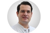
Professor Duška Dragun, Director of the Biomedical Innovation Academy (BIA) of the Berlin Institute of Health (BIH) and Head of the Charité BIH Clinician Scientist Program, succumbed to her long, severe, bravely endured illness on December 28, 2020 at the age of 51.
 
Her tireless efforts were devoted to her life's work: the Charité BIH Clinician Scientist Program. Ten years ago, she launched the first Clinician Scientist Program in Berlin and over the decade established and continuously expanded it as "best practice" for the German-speaking region. She has played a key role in developing and shaping the various programs for scientifically active physicians: from the Clinician Scientist Program, which enables aspiring specialists to spend up to 50 percent of their working time on research, to the Junior Clinician Scientist Program with 20 percent working time on research, which begins in the first year of specialist training, to the Advanced Clinician Scientist Program for specialists with postdoctoral qualifications. Two years ago, she successfully applied to the German Research Foundation (DFG) for the first and only Digital Clinician Scientist Program in Germany. This enables young physicians and scientists to conduct research and work in the field of digitalization in medicine and healthcare. Thus, within a few years, Duška Dragun made a significant contribution to building a new generation of young professionals for medicine – the impact of her programs will last for a long time, via promising individual careers as well as via the programmatic strengthening of a patient-oriented science.  
 
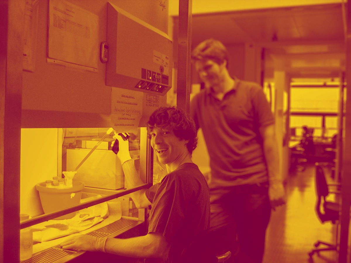
As a physician herself, Professor Duška Dragun has always been committed to research: As acting senior physician and deputy to the acting director of the Department of Nephrology and Intensive Care Medicine at the Charité Campus Virchow-Klinikum, as well as head of the nephrology research laboratory, she made highly regarded, internationally distinguished contributions to transplantation research with the goal of improving graft approach and long-term survival, preventing cardiovascular comorbidity, and thus improving the quality of life and life expectancy of transplanted patients.  She pursued her great goals with tremendous energy and passion, impressive perseverance and clear determination. She devoted her full attention to her employees, colleagues, and students, being equally attentive, understanding, and demanding.
 
The death of Duška Dragun is a great and painful loss. We will miss her greatly as director of the Charité BIH Clinician Scientist Program, as a physician, university professor and scientist. To us she was an inspiration, a mentor and an ever driving force.

Above all, however, we will greatly miss Duška as a friend.  
Karl Hillebrandt | Charité 3R Tandem project for early career researchers
Stacks Image 28699
Together with Dr. Björn Papke (Molecular tumour pathology), Dr. Karl Hillebrandt was able to acquire funding for a "Tandem project for early career researchers" from the Charité 3R. The project is entitled "A personalised therapy approach implementing individually matched matrix-based in vitro colorectal liver metastases to reduce metastatic mouse models".
Although modern multimodal therapy strategies have improved the clinical outcome of patients with colorectal liver metastases (CRLM), the overall prognosis is still poor. To further improve treatment options for patients, it is necessary to develop and test new targeted therapeutic approaches. To date, mouse models have often been used to study metastatic colorectal cancer. However, the rate of successful translation of animal models into clinical trials is less than 8%, highlighting the urgent need for alternative models to study the biology of metastatic cancer. This project aims to develop a novel personalised extracellular matrix-based in vitro model of human CRLM. This model will be validated against existing data from patient-derived organoids and xenografts (histology, single cell RNA sequencing and targeted gene sequencing). After internal comparison of our in vitro CRLM with the original CRLM, we will translate it into a personalised drug screening platform to test drug response from standard therapy to novel inhibitor combinations.
Engineering an endothelialized, endocrine NeoPancreas
Stacks Image 28738
Acta Biomaterialia accepted our latest paper on "Engineering an endothelialized, endocrine Neo-Pancreas: evaluation of islet functionality in an ex vivo model".

Islet-based recellularization of decellularized, repurposed rat livers may form a transplantable Neo-Pancreas. The aim of this study is the establishment of the necessary protocols, the evaluation of the organ structure and the analysis of the islet functionality ex vivo.
After perfusion-based decellularization of rat livers, matrices were repopulated with endothelial cells and mesenchymal stromal cells, incubated for 8 days in a perfusion chamber and finally repopulated on day 9 with intact rodent islets. Integrity and quality of re-endothelialization was assessed by histology and FITC-dextran perfusion assay. Functionality of the islets of Langerhans was determined on day 10 and day 12 via glucose stimulated insulin secretion.
Blood gas analysis variables confirmed the stability of the perfusion cultivation. Histological staining showed that cells formed a monolayer inside the intact vascular structure. These findings were confirmed by electron microscopy. Islets infused via the bile duct could histologically be found in the parenchymal space. Adequate insulin secretion after glucose stimulation after 1-day and 3-day cultivation verified islet viability and functionality after the repopulation process.
We provide the first proof-of-concept for the functionality of islets of Langerhans engrafted in a decellularized rat liver. Furthermore, a re-endothelialization step was implemented to provide implantability. This technique can serve as a bioengineered platform to generate implantable and functional endocrine Neo-Pancreases.

Authors are Hannah Everwien, Eriselda Keshi, Karl H. Hillebrandt, Barbara Ludwig, Marie Weinhart, Peter Tang, Anika S. Beierle, Hendrik Napierala, Joseph MGV Gassner, Nicolai Seiffert, Simon Moosburner, Dominik Geisel, Anja Reutzel-Selke, Benjamin Strücker, Johann Pratschke, Nils Haep, and Igor M. Sauer.
Stacks Image 28740
EKFS grant | Metabolic reconditioning of steatotic rat liver grafts by normothermic ex vivo machine perfusion
Stacks Image 28760
The Else Kröner Fresenius Stiftung will fund the project "Metabolic reconditioning of steatotic rat liver grafts by normothermic ex vivo machine perfusion" (PI: Priv.-Doz. Dr. Nathanael Raschzok) for two years.

Liver transplantation is the treatment of choice for end-stage liver disease, yet the number of transplant candidates constantly exceeds the organ supply. The imbalance between demand and supply of liver grafts is dramatically exacerbated by the rising prevalence of obesity and the metabolic syndrome, which both show a strong correlation with steatosis hepatis. Liver grafts with macrovesicular steatosis above 30% are associated with delayed graft function and lower graft and patient survival, and livers with >60% steatosis are generally discarded from transplantation. Within the next 10 years, the overall liver graft utilization could potentially be halved due to the rising prevalence of steatosis, emphasizing the urgent clinical need to find solutions to make steatotic livers acceptable for transplantation.

In this project the hypothesis is tested whether metabolic reprogramming of steatotic liver grafts will 1) restore hepatocyte function, 2) activate lipid catabolism, 3) increase resistance to ischemia reperfusion damage, and 4) alleviate overwhelming inflammatory processes in the early phase of post-transplant regeneration with beneficial long-term impact for graft function and recipient survival.
The Human Liver Matrisome
Stacks Image 28765
Biomaterials accepted our latest paper on „The Human Liver Matrisome – Proteomic Analysis of Native and Fibrotic Human Liver Extracellular Matrices for Organ Engineering Approaches“.

The production of biomaterials that endow significant morphogenic and microenvironmental cues for the constitution of cell integration and regeneration remains a key challenge in the successful implementation of functional organ replacements. Despite the vast development in the production of biological and architecturally native matrices, the complex compositions and pivotal figures by which the human matrisome mediates many of its essential functions are yet to be defined. Here we present a thorough analysis of the native human liver proteomic landscape using decellularization and defatting protocols to extract create extracellular matrix scaffolds of natural origin that can further be used in both bottom-up and top-down approaches in tissue engineering based organ replacements. Furthermore, by analyzing human liver extracellular matrices in different stages of fibrosis and cirrhosis, we have identified distinct attributes of these tissues that could potentially be exploited therapeutically and thus require further investigation. The general experimental pipeline presented in this study is applicable to any type of tissue and can be widely used for different approaches in regenerative medicine and in the construction of novel biomaterials for organ engineering approaches.

Authors are A. Daneshgar, O. Klein, G. Nebrich, M. Weinhart, P. Tang, A. Arnold, I. Ullah, J. Pohl, S. Moosburner, N. Raschzok, B. Strücker, M. Bahra, J. Pratschke, I.M. Sauer, and K.H. Hillebrandt. The authors acknowledge the support of the Cluster of Excellence Matters of Activity. Image Space Material funded by the Deutsche Forschungsgemeinschaft (DFG, German Research Foundation) under Germany´s Excellence Strategy – EXC 2025.
Stacks Image 28767
<<  Page 2 / 7  >>
Year
© 2025 Prof. Dr. Igor M. Sauer | Charité - Universitätsmedizin Berlin | Disclaimer

This website or its third-party tools use cookies, which are necessary to its functioning and required to achieve the purpose illustrated in the Disclaimer. By closing this banner, scrolling this page, clicking a link or continuing to browse otherwise, you agree to the use of cookies.