Improved cold storage of human hepatocytes
Stacks Image 30009
As a first result of our latest projects concerning the role of miRNA in liver regeneration the American Journal of Physiology - Regulatory, Integrative and Comparative Physiology has accepted our paper "Temporal expression profiles indicate a primary function for microRNA during the peak of DNA replication after rat partial hepatectomy": The liver has the unique capacity to regenerate after surgical resection. However, the regulation of liver regeneration is not completely understood. Recent reports indicate an essential role for small non-coding microRNAs (miRNAs) in the regulation of hepatic development, carcinogenesis, and early regeneration. We hypothesized that miRNAs are critically involved in all phases of liver regeneration after partial hepatectomy. We performed miRNA microarray analyses after 70% partial hepatectomy in rats under isoflurane anesthesia at different time points (0 hours - 5 days) and after sham laparotomy. Putative targets of differentially expressed miRNAs were determined using a bioinformatic approach. 2D-PAGE proteomic analyses and protein identification were performed on specimens at 0 and 24 hours after resection. The temporal dynamics of liver regeneration were characterized by BrdU, PCNA, IL-6, and HGF. We demonstrate that miRNA expression patterns changed during liver regeneration and that these changes were most evident during the peak of DNA replication at 24 hours after resection. Expression of thirteen miRNAs was significantly reduced 12-48 hours after resection (> 25% change), ouf of which downreguation was confirmed in isolated hepatocytes for 6 miRNAs at 24 hours, whereas three miRNAs were significantly upregulated. Proteomic analysis revealed 65 upregulated proteins; among them 23 represent putative targets of the differentially expressed miRNAs. We provide a temporal miRNA expression and proteomic dataset of the regenerating rat liver, which indicates a primary function for miRNA during the peak of DNA replication. These data will assist further functional studies on the role of miRNAs during liver regeneration. Authors are N. Raschzok, W. Werner, H. Sallmon, N. Billecke, C. Dame, P. Neuhaus and I.M. Sauer.
Read More
Profiles of microRNA after rat partial hepatectomy
Stacks Image 30019
The website of the 24th International Congress of The Transplantation Society is online! Please go to www.transplantation2012.org for more information.
Following the exceptionally successful meetings in Boston, Sydney and Vancouver, we would like to welcome you to a city which twenty years after reunification has evolved into one of the most important capitals of Europe. Once the capital of Prussia and leading cultural centre of the 1920s, today the new capital of Germany is characterized by its dazzling modernity and breathtaking architecture. Berlin is young, dynamic and bursting with joie de vivre. Few cities has been shaped to such an extent by history and undergone as much major transformation as Berlin. “Berlin is always in the process of becoming”, remarked historian Karl Scheffler. Even in the 21st century transplantation still is in a process of becoming, too. Therefore, the 24th International Congress of The Transplantation Society will promote the dialogue of experts from around the world. An attractive scientific program will be developed together with the Deutsche Transplantationsgesellschaft (DTG).
The 24th Congress is designed for physicians, surgeons, scientists and organ procurement personnel, who are interested in clinical and research aspects of solid organ, cell and tissue transplantation. The program is developed to encourage the exchange of new scientific and clinical information, and and support an interchange of opinions regarding care and management issues, as well as socioeconomic, ethical and regulatory issues relevant to transplantation.
In addition to the classical types of scientific sessions including plenary sessions, symposia, workshops and poster presentations, we will also offer new types of scientific sessions within the Forum Futurum focusing on Tailored Pharmacotherapy, Imaging and Regenerative Medicine. All of this is designed to develop a highly interactive forum to discuss cutting-edge science in our field.
We therefore cordially invite you and your colleagues together with your friends and family to come to Berlin. Your active participation in the scientific program will be the fundament for a successful event in 2012.
Read More
Monitoring cell transplantation in swine model via MRI

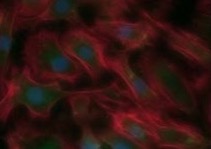
Nora Kammer's paper in Artificial Organs on "Labelling of primary human hepatocytes with micron-sized iron oxide particles in suspension culture suitable for large-scale preparation" is available pre-print. Co-authors are Nils Billecke, Mehmet H. Morgul, Michaela K. Adonopoulou, Martina Mogl, Mao D. Huang, Stefan Florek, Katharina R. L. Schmitt, Nathanael Raschzok and Igor M. Sauer.

Read More
Labelling of hepatocytes in suspension culture
Stacks Image 30053
Our latest paper on "Monitoring of liver cell transplantation in a preclinical swine model using magnetic resonance imaging" has been accepted for publication in CELL Medicine (Part B of CELL TRANSPLANTATION). Authors are Nathanael Raschzok, Ulf Teichgräber, Nils Billecke, Anja Zielinski, Kirsten Steinz, Nora N. Kammer, Mehmet H. Morgul, Sarah Schmeisser, Michaela K. Adonopoulou, Lars Morawietz, Bernhard Hiebl, Ruth Schwartlander, Wolfgang Rüdinger, Bernd Hamm, Peter Neuhaus and Igor M. Sauer. The study was based on the excellent colaboration with the department of Radiology and the Institute of Pathology, both Charité - Campus Mitte, Universitätsmedizin Berlin, Berlin, Germany, the Centre for Biomaterial Development and Berlin-Brandenburg Centre for Regenerative Therapies (BCRT), Institute for Polymer Research, GKSS Research Centre Geesthacht GmbH, Teltow, Germany, the Department of Materials, ETH Zurich, Zurich, C Switzerland, and Cytonet GmbH, Weinheim, Germany.
Liver cell transplantation (LCT) is a promising treatment approach for certain liver diseases, but clinical implementation requires methods for non-invasive follow-up. Labeling with superparamagnetic iron oxide particles can enable the detection of cells with magnetic resonance imaging (MRI). We investigated the feasibility of monitoring transplanted liver cells by MRI in a preclinical swine model and used this approach to evaluate different routes for cell application. Liver cells were isolated from landrace piglets and labeled with micron-sized iron oxide particles (MPIO) in adhesion. Labeled cells (n = 10), native cells (n = 3) or pure particles (n = 4) were transplanted to minipigs via intraportal infusion into the liver, direct injection into the splenic parenchyma, or intra-arterial infusion to the spleen. Recipients were investigated by repeated 3.0 Tesla MRI and computed tomography angiography up to 8 weeks after transplantation. Labeling with MPIO, which are known to have a strong effect on the magnetic field, enabled non-invasive detection of cell aggregates by MRI. Following intraportal application, which is commonly applied for clinical LCT, MRI was able to visualize the microembolization of transplanted cells in the liver that were not detected by conventional imaging modalities. Cells directly injected into the spleen were retained, whereas cell infusions intraarterially into the spleen led to translocation and engraftment of transplanted cells in the liver, with significantly fewer microembolisms compared to intraportal application. These findings demonstrate that MRI can be a valuable tool for non-invasive elucidation of cellular processes of LCT and - if clinically applicable MPIO are available - for monitoring of LCT under clinical conditions. Moreover, the results clarify mechanisms relevant for clinical practice of LCT, suggesting that the intra-arterial route to the spleen deserves further evaluation.
Read More
SPAD in children with acute liver failure
Stacks Image 30063
Hannelore Ringe published the investigations concerning the applicability, efficacy, and safety of single-pass albumin dialysis in children ("Continuous veno-venous single-pass albumin hemodiafiltration in children with acute liver failure." Pediatr Crit Care Med. 2010 Oct 1. [Epub ahead of print]). Co-authors are Varnholt V, Zimmering M, Luck W, Gratopp A, König K, Reich S, Sauer IM, Gaedicke G, and Querfeld U.
The paper reports on a retrospective data review of uncontrolled clinical data of an university-based pediatric intensive care unit collaborating with a local center for liver transplantation. Nine children, aged 2 to 15 yrs, who were treated with single-pass albumin dialysis for acute liver failure of various origins under a compassionate-use protocol between 2000 and 2006. All patients met high-urgency liver transplantation criteria. Single-pass albumin dialysis was performed as rescue therapy for children with acute liver failure. The decrease in hepatic encephalopathy (grades 1-4) and the serum levels of bilirubin, bile acids, and ammonium were measured to assess the efficacy of detoxification. As a measure of liver synthesis function, thromboplastin time and fibrinogen were analyzed. The safety of the procedure was assessed by documenting adverse effects on mean arterial blood pressure, platelet count, and clinical course. Seven out of nine patients were bridged successfully to either native organ recovery (n = 1) or liver transplantation (n = 7), one of them twice. Six out of nine patients undergoing single-pass albumin dialysis (ten treatments) survived. In six patients, hepatic encephalopathy could be reduced at least by one degree. Ammonium, bilirubin, and bile acid levels decreased in all patients. One patient had an allergic reaction to albumin. In childhood acute liver failure, treatment with single-pass albumin dialysis was generally well tolerated and seems to be effective in detoxification and in improving blood pressure, thus stabilizing the critical condition of children before liver transplantation and facilitating bridging to liver transplantation. It may be beneficial in avoiding severe neurologic sequelae after acute liver failure and thereby improve survival. Single-pass albumin dialysis is an inexpensive albumin-based detoxification system that is easy to set up and requires little training. Whether and to what extent single-pass albumin dialysis can support children with acute liver failure until native liver recovery remain unclear.
Read More
Presentations at this year´s XXXVII ESAO Congress
Stacks Image 30073
At this year's XXXVII ESAO Congress, in Skopje. R. Macedonia, Wiebke Werner and Nathanael Raschzok gave oral presentations.
Nathanael Raschzok presented data on "In vitro evaluation of magnetic resonance imaging contrast agents for labeling of human liver cells" (N. Raschzok, D. A. Muecke, M. Adonopoulou, N. Billecke, A. Zielinski, W. Werner, U. Teichgraeber, I. M. Sauer). Wiebke Werner reported on "Temporal microRNA gene expression profiles of the regenerating rat liver after partial hepatectomy" (W. Werner, N. Raschzok, H. Sallmon, N. Billecke, C. Dame, P. Neuhaus, I. M. Sauer), a joint project of the Department of Neonatology, Charité, and our group.
Read More
XXIII International Congress of TTS
Stacks Image 30083
Nathanael Raschzok presented our latest results with labeled hepatocytes in the pig model at the XXIII International Congress of The Transplantation Society in Vancouver, Canada. The oral presentation was entitled "MRI enables monitoring of transplanted hepatocytes in a preclinical large animal model" (N. Raschzok, N. Billecke, A. Zielinski, K. Steinz, N.N. Kammer, S. Schmeisser, M.H. Morgul, M.K. Adonopoulou, J. Pinkernelle, L. Morawietz, B. Hiebl, W. Rüdinger, U. Teichgräber, P. Neuhaus, I.M. Sauer).
Read More
Hypothermia-induced cell protection & microglial cells
Stacks Image 30100
The European Journal of Neuroscience published the paper "Mechanisms of hypothermia-induced cell protection mediated by microglial cells in vitro" First author is Antje Diestel from Katharina Schmitt's group - co-authors are Silke Troeller, Nils Billecke, Igor M. Sauer, Felix Berger and Katharina R. L. Schmitt.
Despite the widespread interest in the clinical applications of hypothermia, the cellular mechanisms of hypothermia-induced neuroprotection have not yet been clearly understood. Therefore, the aim of this study was to elucidate the cellular effects of clinically relevant hypothermia and rewarming on the morphological and functional characteristics of microglia. Microglial cells were exposed to a dynamic cooling and rewarming protocol. For stimulation, microglial cells were treated with 1 μg/mL lipopolysaccharide (LPS). We found that hypothermia led to morphological changes from ramified to ameboid cell shapes. At 2 h after hypothermia and rewarming, microglial cells were again ramified with extended branches. Moreover, we found enhanced cell activation after rewarming, accompanied by increased phagocytosis and adenosine triphosphate consumption. Interestingly, hypothermia and rewarming led to a time-dependent significant up-regulation of the anti-inflammatory cytokines interleukin-10 and interleukin-1 receptor antagonist in stimulated microglial cells. This is in line with the reduced proliferation and time-dependent down-regulation of the pro-inflammatory cytokines tumor necrosis factor-alpha and monocyte chemotactic protein-1 in comparison to normothermic control cells after LPS stimulation. Furthermore, degradation of the inhibitor of the nuclear transcription factor-kappaB (IkappaB-alpha) was diminished and delayed under conditions of cooling and rewarming in LPS-stimulated microglial cells. Thus, our results show that hypothermia and rewarming activate microglial cells, increase phagocytosis and shift the balance of cytokine release in stimulated microglial cells towards the anti-inflammatory cytokines. This could be a new cellular mechanism of hypothermia-induced neuroprotection mediated by activated microglial cells.
European Journal of Neuroscience, 2010; 31: 779-787
Read More
Modified nanoparticles & multimodal imaging
Stacks Image 30117
Lars Stelter's studies on In vitro and in vivo detectability of modified superparamagnetic nanoparticles for multimodal imaging using fluorescence microscopy, 3T MRI and animal PET are published in the latest issue of Molecular Imaging & Biology (Mol Imaging Biol. 2010 Jan-Feb;12(1):25-34). Co-authors are Jens Pinkernelle, Roger Michel, Ruth Schwartländer, Nathanael Raschzok, Mehmet H. Morgul, Martin Koch, Timm Denecke, Holger Amthauer, Juri Ruf, Andreas Jordan, Bernd Hamm, Igor M. Sauer, Ulf Teichgräber.
Cell transplantation is a major field in regenerative medicine and a promising alternative to whole organ transplantation. However, the process of cell engraftment is not yet fully understood and the hitherto achieved clinical outcome is limited. The aim of our study was to modify an aminosilan-coated nanoparticle for cell labeling and make it applicable for multimodal imaging using MRI, PET and fluorescent imaging. HIV-1 tat, linked FITC, and Gallium-68 were covalently bound to the particle and injected into Wistar rats. Animal-PET imaging was performed followed by MRI at 3.0T. Hepatic accumulation of the particles was proven by radionuclide distribution after 10 minutes in PET as well as in MRI over a 24 hour-period. Histological workup of the liver also revealed content of iron oxide particles in the reticuloendothelial system. Adjacent in vitro studies incubating hepatogenic HuH7 cells with the particles showed a rapid intracellular accumulation, clearly detectable by fluorescence microscopy and MRI. In conclusion our modified nanoparticle is stable under in vitro and in vivo conditions and is applicable for multimodal molecular imaging. Cellular labeling with this particle is possible and might help to get new insights into understanding the process of cell transplantation.
Read More
XXIII International Congress of TTS
Stacks Image 30122

The XXIII International Congress of The Transplantation Society, the leading international congress on transplantation biology and medicine, will be held in Vancouver, Canada from 15 - 19 August 2010. The translational program will draw together experts from around the world, and will encompass innovations in genomics and proteomics, molecular analyses of human diseases, innovations in biological and pharmacological immunosuppression, novel approaches to tolerance induction, technical advances in robotic surgery and imaging, advances in clinical practice and many other topics at the cutting edge of stem-cell, cellular and solid organ transplantation.
The XXIII Congress will be one of the largest and most exciting international gatherings of clinicians and scientists in the field of transplantation, drawing more than 4,000 delegates from all parts of the world. The international delegation will enjoy a stellar Scientific Program, commencing with a comprehensive and cutting-edge Postgraduate Education Program on Saturday 14th August and Sunday 15th August, and continuing with a panoply of outstanding plenary symposia, state of the art lectures, individual scientific presentations, and thematic symposia from Monday 16th August to Thursday 19th August inclusive.
The 2010 Congress will provide an outstanding opportunity for networking with colleagues and leading experts, and for sharing and discussing clinical advances, late-breaking discoveries and therapeutic advances in this field.
More information is available via www.transplantation2010.org. Furthermore, you may download the 2nd announcement here.
Important dates:
Abstract Submission Opens: October 2009
Online Registration Opens: November 2009
Abstract Submission Deadline: 5 March 2010
Author Notification of Acceptance: 7 May 2010
Early Registration Deadline: 4 June 2010
Late Breaking Abstract Deadline: 11 June 2010
Hotel Reservation Deadline: 25 June 2010
Regular Registration Deadline: 16 July 2010

Read More
Haluk Morgül defended thesis "magna cum laude"
Haluk Morgül successfully defended his medical doctoral thesis "magna cum laude".
After years of extremely fruitful research in the field of liver support, hepatocyte isolation and cell imaging via MRI he is (co-)author of 5 papers in peer reviewed journals (with more to come)!
Read More
cBAL111
Stacks Image 30143
Tanja Deurholt's paper on "Novel immortalized human fetal liver cell line, cBAL111, has the potential to differentiate into functional hepatocytes" is published in BMC Biotechnology. Co-authors are Niek P. van Til, Aniska A. Chhatta, Lysbeth ten Bloemendaal, Ruth Schwartlander, Catherine Payne, John N. Plevris, Igor M. Sauer, Robert A.F.M. Chamuleau, Ronald P.J. Oude Elferink, Jurgen Seppen, and Ruurdtje Hoekstra.
A clonal cell line that combines both stable hepatic function and proliferation capacity is desirable for in vitro applications that depend on hepatic function, such as pharmacological or toxicological assays and bioartificial liver systems. The article describes the generation and characterization of a clonal human cell line for in vitro hepatocyte applications.Cell clones derived from human fetal liver cells were immortalized by over-expression of telomerase reverse transcriptase. The resulting cell line, cBAL111, displayed hepatic functionality similar to the parental cells prior to immortalization, and did not grow in soft agar. Cell line cBAL111 expressed markers of immature hepatocytes, like glutathione S transferase and cytokeratin 19, as well as progenitor cell marker CD146 and was negative for lidocaine elimination. On the other hand, the cBAL111 cells produced urea, albumin and cytokeratin 18 and eliminated galactose. In contrast to hepatic cell lines NKNT-3 and HepG2, all hepatic functions were expressed in cBAL111, although there was considerable variation in their levels compared with primary mature hepatocytes. When transplanted in the spleen of immunodeficient mice, cBAL111 engrafted into the liver and partly differentiated into hepatocytes showing expression of human albumin and carbamoylphosphate synthetase without signs of cell fusion. This novel liver cell line has the potential to differentiate into mature hepatocytes to be used for in vitro hepatocyte applications.
Read More
ESAO 2009 - presentations
At the XXXVI Annual Meeting of the European Society for Artificial Organs ESAO 2009 in Compiègne/France the following projects have been presented by members of our group:

Tracking of transplanted liver cells via clinical 3.0 tesla MRI A. Zielinski, K. Steinz, N. Raschzok, N. Billecke, N. Kammer, M.H. Morgul, M. Adonopulou, S. Schmeisser, J. Pinkernelle, W. Ruedinger, U. Teichgraeber, I.M. Sauer
Evaluation of application sites for liver cell transplantation in a large animal model K. Steinz, A. Zielinski, N. Raschzok, N. Billecke, N.N. Kammer, M.H. Morgul, M. Adonopulou, L. Morawietz, W. Rüdinger, I.M. Sauer
Methylprednisolone and tacrolimus prevent hypothermia induced endothelial dysfunction - possible use in transplantation surgery K.R.L. Schmitt, A. Diestel, N. Billecke, F. Berger, I.M. Sauer
Read More
One Day of the Liver 2009
Stacks Image 30176
The traditional One Day on the Liver will be part of the XXXVI Annual Meeting of the European Society for Artificial Organs ESAO 2009 at the Université de Technologie de Compiègne in France. ODOL 2009 will take place September, 3rd focussing on "Cells, Hybrids, and Machines".
The following keynote lectures will be given:
Clinical Results of Liver Cell Therapy in Children with Urea Cycle Defect by Dr. Dr. W. Rüdinger, Weinheim, Germany,
Xenotransplantation. Where are we in 2009 ? by Dr. A. Billiau, Leuven, Belgium,
Problems and Opportunities: Perspectives of Bioartificial Liver Support by Dr. R. Hoekstra, Amsterdam, Netherlands,
New perspectives of artificial liver support by Prof. Dr. D. Falkenhagen, Krems, Austria.

We expect to present you all lectures as video streams after the meeting.
Read More
HAI 2009 - New aspects in liver support
Stacks Image 30186
The group was awarded with a research bonus (Forschungsprämie) by the Bundesministeriums für Bildung und Forschung (BMBF, Federal Ministry of Education and Research) supporting the development of micron sized particles for the detection of transplanted cells via MRI. The program is part of the High-Tech Strategy for Germany, launched by the Federal Government in August 2006 to encourage the development of new products and innovative services.
Read More
Nathanael Raschzok defended thesis "summa cum laude"
Stacks Image 30196
Nathanael Raschzok successfully defended his medical doctoral thesis "summa cum laude". After three years of extremely fruitful research and development in the field of hepatocyte transplantation, cell labeling, and MR imaging of transplanted cells he is first author of three papers in peer reviewed journals (with more to come...). He currently is finishing his in vivo MRI studies of MPIO labeled transplanted hepatocytes.
Transplantation of primary human hepatocytes is a promising approach in certain liver diseases. For visualisation of hepatocytes during and following cell application and the ability of a timely response to potential complications, a non-invasive modality for imaging of the transplanted cells has to be established. The aim of his studies was to label primary human hepatocytes with micron-sized iron oxide particles (MPIOs), enabling the detection of cells by clinical magnetic resonance imaging (MRI). The feasibility of preparing MPIO-labelled primary human hepatocytes detectable by clinical MR equipment was shown in vitro. MPIO-labelled cells could serve for basic research and quality control in the clinical setting of human hepatocyte transplantation.
He also investigated techniques for evaluation of the particle uptake via continuum source atomic absorption spectrometry (CSAAS). Porcine liver cells were labelled with MPIOs and the iron concentration of the cell samples was investigated by a CSAAS spectrometer equipped with a Perkin-Elmer THGA graphite furnace. CSAAS enabled rapid quantification of particle load from small quantities of cells without extensive preparation steps. CSAAS could be used for quality control in a clinical setting of cell transplantation.
Read More
Hypothermia induced endothelial dysfunction
Stacks Image 30206
Antje Diestel's manuscript entitled "Tacrolimus and methylprednisolone prevent hypothermia induced endothelial dysfunction" has been accepted for publication in the Journal of Heart and Lung Transplantation. Co-authors are Nils Billecke, Joerg Roessler, Boris Schmitt, Silke Troeller, Ruth Schwartlander, Felix Berger, Igor Maximilian Sauer and Katharina Rose Luise Schmitt.
Hypothermia is used to preserve organs for transplant and it is the oldest method to protect organs during complex pediatric cardiac surgery. Loss of tissue function and tissue edema are common complications in children undergoing cardiac surgery and heart transplantation. The present study was designed to examine the effects of methylprednisolone (MP) and Tacrolimus (TAC) on endothelial cell function and morphology after deep hypothermia and rewarming. Human umbilical vein endothelial cells (HUVECs) were pretreated with MP and/orTAC and incubated either within a specially designed bioreactor or in monolayers. They were then exposed to a dynamic cooling and rewarming protocol. Immunocytochemistry, time lapse video microscopy within the SlideReactor bioreactor system, cell permeability and adherence assays and western blot analysis were performed. Confluent endothelial cells exposed to hypothermia displayed elongated cell shapes with intercellular gap formation, increased endothelial cell-layer permeability and loss in adherence. Upon rewarming, however, endothelial cell integrity was restored. Opening and closing of intercellular gaps was dependent on ERK 1/2 activation and connexin 43 (Cx43) expression. The combined treatment with MP and TAC inhibited these hypothermia-induced changes. These results suggest that MP and TAC inhibit hypothermia induced endothelial gap formation via pERK 1/2 inhibition and connexin 43 stabilization. Application of combined drugs that affect multiple targets may therefore be considered as a possible new therapeutic strategy to prevent endothelial dysfunction after hypothermia and rewarming.
Read More
CSAAS and MPIO-labelled cells
Stacks Image 30216
As a result of the fruitful collaboration with the Institute for Analytical Sciences Berlin Nathanael Raschzok's paper on "Quantification of Cell Labelling with Micron-Sized Iron Oxide Particles Using Continuum Source Atomic Absorption Spectrometry" has been accepted by Tissue Engineering for publication. Co-authors are Nils Billecke, Nora N. Kammer, Mehmet H. Morgul, Michaela K. Adonopoulou, Igor M. Sauer, Stefan Florek, Helmut Becker-Ross, and Mao-Dong Huang.

Detection of cells after transplantation is necessary for quality control in regenerative medicine. Labelling with micron-sized iron oxide particles (MPIOs) enables non-invasive detection of single cells by magnetic resonance imaging. However, techniques for evaluation of the particle uptake are challenging. The aim of this study was to investigate continuum source atomic absorption spectrometry (CSAAS) for this purpose. Porcine liver cells were labelled with MPIOs and the iron concentration of the cell samples was investigated by a CSAAS spectrometer equipped with a Perkin-Elmer THGA graphite furnace. The weak iron line at 305.754 nm provides only about 1/600 sensitivity of the iron resonance line at 248.327 nm and was used for CSAAS measurements. Iron concentrations measured from labelled cells ranged from (5.8 ± 0.3) to (25.8 ± 0.9) pg Fe/cell, correlating to an uptake of (8.2 ± 0.5) to (25.7 ± 0.8) particles/cell. The results were verified by standardised morphometric evaluation. CSAAS enabled rapid quantification of particle load from small quantities of cells without extensive preparation steps. Thereby, CSAAS could be used for quality control in a clinical setting of cell transplantation.
Read More
Dr. med. Dominik Paul Modest
After years of research in the field of hepatocyte isolation and hypothermic long-term culture Dominik Paul Modest successfully defended his thesis "magna cum laude".
Read More
Dr. Igor M. Sauer: obtained postdoctoral lecture qualification (Habilitation) in Surgery
February 5th, 2009 at 4 p.m. (Hörsaal 3, Lehrgebäude Charité - Campus Virchow Klinikum) Dr. I.M. Sauer will give his inaugurative lecture entitled "Künstliche Organe: Von der Vision kybernetischer Organismen zur medizinischen Realität".
Read More
Imaging of human hepatocytes via MPIO and MRI
Stacks Image 30278
Nathanel Raschzok's and Haluk Morgül's manuscript entitled "Imaging of Primary Human Hepatocytes Using Micron-Sized Iron Oxide Particles and Clinical Magnetic Resonance Tomography" has been accepted for publication in the Journal of Cellular and Molecular Medicine (impact factor: 6,55). Authors are Nathanael Raschzok, Mehmet H. Morgul, Jens Pinkernelle, Florian W.R. Vondran, Nils Billecke, Nora N. Kammer, Gesine Pless, Michaela K. Adonopoulou, Christian Leist, Lars Stelter, Ulf Teichgraber, Ruth Schwartlander and Igor M. Sauer. Nathanael Raschzok and Mehmet Haluk Morgul contributed equally to this work. The contribution of Ruth Schwartländer has to be emphasised as well. Transplantation of primary human hepatocytes is a promising approach in certain liver diseases. For visualisation of hepatocytes during and following cell application and the ability of a timely response to potential complications, a non-invasive modality for imaging of the transplanted cells has to be established. The aim of this study was to label primary human hepatocytes with micron-sized iron oxide particles (MPIOs), enabling the detection of cells by clinical magnetic resonance imaging (MRI). Primary human hepatocytes isolated from 13 different donors were used for labelling experiments. Following dose finding studies, hepatocytes were incubated with 30 particles/cell for 4 hours in adhesion culture. Particle incorporation was investigated via light, fluorescence and electron microscopy and labelled cells were fixed and analysed in an agarose suspension by a 3.0 Tesla MR scanner. Hepatocytes were enzymatically resuspended and analysed during a five-day reculture period for viability, total protein, enzyme leakage (AST, LDH) and metabolic activity (urea, albumin). A mean uptake of 18 particles/cell could be observed, and primary human hepatocytes were clearly detectable by MR instrumentation. The particle load was not affected by resuspension and showed no alternations during the culture period. Compared to control groups, labelling and resuspension had no adverse effects on viability, enzyme leakage and metabolic activity of human hepatocytes. Conclusion: The feasibility of preparing MPIO-labelled primary human hepatocytes detectable by clinical MR equipment was shown in vitro. MPIO-labelled cells could serve for basic research and quality control in the clinical setting of human hepatocyte transplantation.
Read More
Tat-peptide modified MPIO
Stacks Image 30288
Haluk Morgül and Nathanael Raschzok published their first results on "Tracking of primary human hepatocytes with clinical MRI: Initial results with Tat-peptide modified superparamagnetic iron oxide particles." in the March issue of IJAO (Int J Artif Organs 2008, 31:252-257): The transplantation of primary human hepatocytes is a promising approach in the treatment of specific liver diseases. However, little is known about the fate of the cells following application. Magnetic resonance imaging (MRI) could enable real-time tracking and long-term detection of transplanted hepatocytes. The use of superparamagnetic iron oxide particles as cellular contrast agents should allow for the non-invasive detection of labelled cells on high-resolution magnetic resonance images. Experiments were performed on primary human hepatocytes to transfer the method of detecting labelled cells via clinical MRI into human hepatocyte transplantation. For labelling, Tat-peptide modified nano-sized superparamagnetic MagForce particles were used. Cells were investigated via a clinical MR scanner at 3.0 Tesla and the particle uptake within single hepatocytes was estimated using microscopic examinations. The labelled primary human hepatocytes were clearly detectable by MRI, proving the feasibility of this new concept. Therefore, this method is a useful tool to investigate the effects of human hepatocyte transplantation and to improve safety aspects of this method.
Read More
<<  Page 7 / 7 
Year
© 2025 Prof. Dr. Igor M. Sauer | Charité - Universitätsmedizin Berlin | Disclaimer

This website or its third-party tools use cookies, which are necessary to its functioning and required to achieve the purpose illustrated in the Disclaimer. By closing this banner, scrolling this page, clicking a link or continuing to browse otherwise, you agree to the use of cookies.