Brigitta Globke: Digital Clinician Scientist
Stacks Image 28789
Dr. Brigitta Globke successfully applied for participation in the BIH Charite Digital Clinician Scientist Program.

The aim of the project is the development and evaluation of an augmented reality assist system for intraoperative photoplethysmographic control of perfusion. The project is carried out in collaboration with Benjamin Kossack, Fraunhofer | Heinrich Hertz Institute Computer Vision and Graphics.

Charité and BIH are jointly organizing the new "Digital Clinician Scientist Program" (D-CSP). The program is primarily aimed at physicians who are already working on innovative research projects to meet the technological challenges of data-driven medicine during their specialist training. The German Research Foundation (DFG) is funding the project for an initial period of three years.

The BIH Charité Digital Clinician Scientist Program will provide a new career path for the creators of digital change in medicine and will expand the successful Germany-wide model of the BIH Charité Clinician Scientist Program. In addition to the three-year individual funding, which is based on protected time for research, the focus is on modules for the acquisition of scientific skills (Big Data, bioinformatics or artificial intelligence) as well as mandatory mentoring. For the new program, various experts* from the Charité, the BIH, the Max Delbrück Center for Molecular Medicine (MDC), the Berlin Institute for Medical Systems Biology (BIMSB), the Einstein Center for Digital Future, and the Bernstein Center for Computational Neuroscience will be involved in the design of the D-CSP and in the recruitment and supervision of program participants.
Dual versus single vessel normothermic ex vivo perfusion of rat liver grafts using metamizole for vasodilatation
Stacks Image 28794
F. Claussen, J.M.G.V. Gassner, S. Moosburner, D. Wyrwal, M. Nösser, P. Tang, L. Wegener, J. Pohl, A. Reutzel-Selke, R. Arsenic, J. Pratschke, I.M. Sauer, and N. Raschzok published their trecent work on "Dual versus single vessel normothermic ex vivo perfusion of rat liver grafts using metamizole for vasodilatation" in PLoS One 2020;15(7): e0235635.

Normothermic ex vivo liver perfusion (NEVLP) is a promising strategy to increase the donor pool in liver transplantation. Small animal models are essential to further investigate questions regarding organ preservation and reconditioning by NEVLP. A dual vessel small animal NEVLP (dNEVLP) model was developed using metamizole as a vasodilator and compared to conventional portovenous single vessel NEVLP (sNEVLP).

Livers of male Wistar rats were perfused with erythrocyte-supplemented culture medium for six hours by either dNEVLP via hepatic artery and portal vein or portovenous sNEVLP. dNEVLP was performed either with or without metamizole treatment. Perfusion pressure and flow rates were constantly monitored. Transaminase levels were determined in the perfusate at the start and after three and six hours of perfusion. Bile secretion was monitored and bile LDH and GGT levels were measured hourly. Histopathological analysis was performed using liver and bile duct tissue samples after perfusion.

Hepatic artery pressure was significantly lower in dNEVLP with metamizole administration. Compared to sNEVLP, dNEVLP with metamizole treatment showed higher bile production, lower levels of transaminases during and after perfusion as well as significantly lower necrosis in liver and bile duct tissue. Biochemical markers of bile duct injury showed the same trend.

Our miniaturized dNEVLP system enables normothermic dual vessel rat liver perfusion. The administration of metamizole effectively ameliorates arterial vasospasm allowing for six hours of dNEVLP, with superior outcome compared to sNEVLP.

Development of GelMA/PCL and dECM/PCL resins for 3D printing of acellular in vitro tissue scaffolds by stereolithography
Stacks Image 28799
Gelatin methacryloyl (GelMA) is a chemically modified extracellular matrix (ECM)-derived biopolymer that is widely used for 3D fabrication of tissue engineering scaffolds. However, its tendency for physical gelation limits its use in aqueous 3D printing resins to low concentrations, yielding a poor printing resolution in stereolithography (SLA).
To obtain a GelMA-based resin that can be printed into high-resolution tissue scaffolds, we formulated resins of fish and porcine-derived GelMA in formamide using GelMA alone or mixed with star-shaped poly(ε-caprolactone) methacrylate (PCL-MA). We identified GelMA resins and GelMA/PCL-MA hybrid resins with a ratio of 70/30 wt-% to yield a suitable viscosity for SLA at 32 °C and demonstrated the resolution of the new resins in SLA by 3D printing acellular human small intestine-mimicking tissue scaffolds. The presence of PCL-MA in the hybrid resins improved the 3D printing fidelity compared to the neat GelMA resins, while GelMA provided the hybrid materials with enhanced swelling and proliferation of seeded cells. We further demonstrated the transferability of our resin formulation to native organ-derived materials by successfully replacing GelMA in the hybrid resin with solubilized, methacryloyl-functionalized decellularized liver ECM (dECM-MA) and by 3D printing multi-layer dECM/PCL-MA hydrogels.

"Development of GelMA/PCL and dECM/PCL resins for 3D printing of acellular in vitro tissue scaffolds by stereolithography" was published in Mater Sci Eng C Mater Biol Appl. 2020 Jul;112:110958. Authors are L. Elomaa, E. Keshi, I.M. Sauer, and M. Weinhart.
Extended reality technologies for support of surgical workflows
Stacks Image 28806
Current developments in the field of extended reality (XR) could prove useful in the optimization of surgical workflows, time effectiveness and postoperative outcome. Although still primarily a subject of research, the state of XR technologies is rapidly improving and approaching feasibility for a broad clinical application. Surgical fields of application of XR technologies are currently primarily training, preoperative planning and intraoperative assistance. For all three areas, products already exist (some clinically approved) and technical feasibility studies have been conducted. In teaching, the use of XR can already be assessed as fundamentally practical and meaningful but still needs to be evaluated in large multicenter studies. In preoperative planning XR can also offer advantages, although technical limitations often impede routine use; however, for cases of intraoperative use informative evaluation studies are mostly lacking, so that an assessment is not yet possible in a meaningful way. Furthermore, there is a lack of assessments regarding cost-effectiveness in all three areas. The XR technologies enable proven advantages in surgical workflows despite the lack of high-quality evaluation with respect to the practical and clinical use of XR. New concepts for effective interaction with XR media also need to be developed. In the future, further research progress and technical developments in the field can be expected.

Authors are Christoph Rüger, Simon Moosburner and Igor M. Sauer (Chirurg 2020; 91(7): 544-552).
Junior Professorship for Digital Surgery and Interdisciplinary Technology Research
Stacks Image 28811
The Department of Surgery of the Charité (Director: Prof. Dr. Johann Pratschke) at the Charité Center 8 (CharitéCenter for Surgery) invites applications for the position of the Junior Professorship for Digital Surgery and interdisciplinary Technology Research (Salary Group: W1 BBesG-ÜfBE, non-tenured) with the reference number: Prof. 546/2020.

The initial appointment is for three years with the optional extension for another three years follow-ing successful evaluation. It is aimed to turn the Junior Professorship into a W2-Professorship (Salary Group: W2 BBesG-ÜfBE) after six years.The successful candidate has to fulfill the appointment requirements in accordance with § 102a of the Berlin Higher Education Act (Berliner Hochschulgesetz, Gem. § 102a BerlHG) and needs to credibly demonstrate through his/her previous scientific work that he/she is able to fulfill the expectations of the junior professorship.

One of the tasks of this Junior Professorship is the appropriate representation of the research area mentioned above. Within the framework of the Cluster of Excellence Matters of Activity – Image Space Material, he/she is expected to evaluate, accompany and advance the digital transformation in surgery and related disciplines as well as expand the repertoire of methods and initiate innovations. In cooperation with the research areas Cutting and Material Form Function of the Cluster of Excellence, new surgical cutting techniques are to be investigated and developed. It is planned to be linked to the currently being established institutions, The Simulated Human Being (Si-M) and the Berlin Simulation and Training Centre (BeST). In addition to the tasks mentioned, the following three fields of activity are to be covered:

Interdisciplinary Knowledge Transfer

  • Implementation of new applications from areas such as deep learning, extended reality (mixed and virtual reality) or robotics in surgical practice requires an intensification of interdisciplinary cooperation
  • Continuous exchange between industry and practice as well as with adjacent disciplines (e.g. Radiology)
  • Integration of a growing number of applications and competencies from areas outside established medical technology, e.g.game design, computer science or human factor studies

Technology Assessment

  • Sustainable implementation of digital technologies through opportunity and risk assessment
  • Advising the Department of Surgery on investment decisions through appropriate risk and media competency
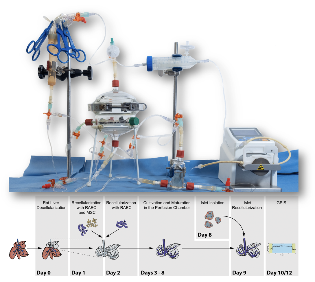
Innovation

  • Identification of concrete application locations and practices of digital surgery within the clinic and experimental research (e.g. use of technologies in the context of biomedical research approaches to organ replacement as well as oncological models) for future Living Labs and to demonstrate these to the public
  • Integration of users, research projects and start-ups also outside the Clinic

The successful candidate will be engaged in teaching activities of the medical education curriculum at Charité, supervise Master and Doctoral candidates, and participate in academic self-organization. In addition, the candidate should present concepts for a good supervision of doctoral students as well as for the integration of his/her research activities into the teaching of the Charité. Appointment requirements are governed by article 102a of the Berlin Higher Education Act (Berliner Hochschulgesetz:§ 102a BerlHG). Completed university degree in Natural Sciences, Humanities and/or Life Sciences or any other related field of Medicine or non-medicine is required. In addition, a Doctorate (Ph.D and/or M.D.) and significant post-doctoral experience are required. Basic medical knowledge is desired.

The Charité is an equal opportunity employer committed to excellence through diversity. As women are under-represented in academics, we explicitly encourage women to send in their application. Women will be given preference over equally qualified men (within the framework of the legal possibilities). We value diversity and therefore welcome all applications – regardless of gender, nationality, social background, religion or age. Equally qualified applicants with disabilities will be given preference.

Written applications according to the format specified on https://career.charite.de/am/calls/application_notes.pdf should be submittedby June 19th, 2020 under https://career.charite.de. For further questions on details, please contact Prof. Dr. Igor Maximilian Sauer.
Magnetic resonance elastography quantification of decellularized liver tissue
Stacks Image 28816
"Magnetic resonance elastography quantification of the solid-to-fluid transition of liver tissue due to decellularization" was published in the latest issue of the Journal of the Mechanical Behavior of Biomedical Materials.

Maintenance of tissue extracellular matrix (ECM) and its biomechanical properties for tissue engineering is one of the substantial challenges in the field of decellularization and recellularization. Preservation of the organ-specific biomatrix is crucial for successful recellularization to support cell survival, proliferation, and functionality. However, understanding ECM properties with and without its inhabiting cells as well as the transition between the two states lacks appropriate test methods capable of quantifying bulk viscoelastic parameters in soft tissues.
We used compact magnetic resonance elastography (MRE) with 400, 500, and 600 Hz driving frequency to investigate rat liver specimens for quantification of viscoelastic property changes resulting from decellularization. Tissue structures in native and decellularized livers were characterized by collagen and elastin quantification, histological analysis, and scanning electron microscopy.
Decellularization did not affect the integrity of microanatomy and structural composition of liver ECM but was found to be associated with increases in the relative amounts of collagen by 83-fold (37.4 ± 17.5 vs. 0.5 ± 0.01 μg/mg, p = 0.0002) and elastin by approx. 3-fold (404.1 ± 139.6 vs. 151.0 ± 132.3 μg/mg, p = 0.0046). Decellularization reduced storage modulus by approx. 9-fold (from 4.9 ± 0.8 kPa to 0.5 ± 0.5 kPa, p < 0.0001) and loss modulus by approx. 7-fold (3.6 kPa to 0.5 kPa, p < 0.0001), indicating a marked loss of global tissue rigidity as well as a property shift from solid towards more fluid tissue behavior (p = 0.0097).
Our results suggest that the rigidity of liver tissue is largely determined by cellular components, which are replaced by fluid-filled spaces when cells are removed. This leads to an overall increase in tissue fluidity and a viscous drag within the relatively sparse remaining ECM. Compact MRE is an excellent tool for quantifying the mechanical properties of decellularized biological tissue and a promising candidate for useful applications in tissue engineering.

Authors are Hannah Everwien, Angela Ariza de Schellenberger, Nils Haep, Heiko Tzschätzsch, Johann Pratschke, Igor M. Sauer, Jürgen Braun, Karl H. Hillebrandt and Ingolf Sack.

J Mech Behav Biomed Mater. 2020 Apr;104:103640. doi: 10.1016/j.jmbbm.2020.103640. Epub 2020 Jan 14.
Characterization of pancreatic and biliary cancer stem cells in patient-derived tissue
Stacks Image 28821
Pancreatic ductal adenocarcinoma (PDAC) and extrahepatic cholangio-carcinoma (eCC) represent two cancer entities with devastating prognoses. Despite recent progress in research and treatment, therapy remains challenging. Cancer stem cells (CSCs) have been shown to play an important role in metastasis and chemoresistance. Therefore, CSCs may play a promising role as a potential therapeutic target.
A total of 31 patients (23 PDAC, 8 eCC) were included in the study. CSCs were analyzed in a single-cell suspension of tumor samples via fluorescence-activated cell scanning (FACS) with a functional Hoechst 33342 staining as well as a cell surface marker staining of the CSC-panel (CD24, CD44 and EpCAM) and markers to identify fibroblasts, leukocytes and components of the notch signaling pathway. Furthermore, the potential presence of CSCs among primary cancer-associated fibroblasts (CAFs) was assessed using the same FACS-panel.
We showed that CSCs are present in patient-derived dissociated tumor tissue. The functional and surface marker profile of CSC-detection did in fact correlate. The amount of CSCs was significantly correlated with tumor characteristics such as a higher UICC stadium and nodal invasion. CSCs were not restricted to the epithelial cell fraction in tumor tissues, which has been verified in independent analysis of primary cell cultures of CAFs.
Our study confirms the in vivo presence of CSCs in PDAC and eCC, stating a clinical significance thereof and thus their plausibility as therapeutic targets. In addition, stem-like cells also seem to constitute a part of the CAFs.

"Characterization of Pancreatic and Biliary Cancer Stem Cells in Patient-derived Tissue" was published in Anticancer Research. Authors are J. Gogolok, E. Seidel, A. Strönisch, A. Reutzel-Selke, I.M. Sauer, J. Pratschke, M. Bahra, and R.B. Schmuck.
Hepatocyte transplantation to the liver via the splenic artery
Stacks Image 28826
Hepatocyte transplantation (HcTx) is a promising approach for the treatment of metabolic diseases in newborns and children. The most common application route is the portal vein, which is difficult to access in the newborn. Transfemoral access to the splenic artery for HcTx has been evaluated in adults, with trials suggesting hepatocyte translocation from the spleen to the liver with a reduced risk for thromboembolic complications. Using juvenile Göttingen minipigs, we aimed to evaluate feasibility of hepatocyte transplantation by transfemoral splenic artery catheterization, while providing insight on engraftment, translocation, viability, and thromboembolic complications. Four Göttingen Minipigs weighing 5.6 kg to 12.6 kg were infused with human hepatocytes (two infusions per cycle, 1.00E08 cells per kg body weight). Immunosuppression consisted of tacrolimus and prednisolone. The animals were sacrificed directly after cell infusion (n=2), 2 days (n=1), or 14 days after infusion (n=1). The splenic and portal venous blood flow was controlled via color-coded Doppler sonography. Computed tomography was performed on days 6 and 18 after the first infusion. Tissue samples were stained in search of human hepatocytes. Catheter placement was feasible in all cases without procedure-associated complications. Repetitive cell transplantations were possible without serious adverse effects associated with hepatocyte transplantation. Immunohistochemical staining has proven cell relocation to the portal venous system and liver parenchyma. However, cells were neither present in the liver nor the spleen 18 days after HcTx. Immunological analyses showed a response of the adaptive immune system to the human cells. We show that interventional cell application via the femoral artery is feasible in a juvenile large animal model of HcTx. Moreover, cells are able to pass through the spleen to relocate in the liver after splenic artery infusion. Further studies are necessary to compare this approach with umbilical or transhepatic hepatocyte administration.

"Hepatocyte Transplantation to the Liver via the Splenic Artery in a Juvenile Large Animal Model" was published in Cell Transplantation.
Authors are J. Siefert, K.H. Hillebrandt, S. Moosburner, P. Podrabsky, D. Geisel, T. Denecke, J.K. Unger, B. Sawitzki, S. Gül-Klein, S. Lippert, P. Tang, A. Reutzel-Selke, M.H. Morgul, A.W. Reske, S. Kafert-Kasting, W. Rüdinger, J. Oetvoes, J. Pratschke, I.M. Sauer, and N. Raschzok.
New book: Decellularized Extracellular Matrix: Characterization, Fabrication and Applications
Stacks Image 28831
The extracellular matrix (ECM) supports cells and regulates various cellular functions in our body. The native ECM promises to provide an excellent scaffold for regenerative medicine. In order to use the ECM as a scaffold in medicine, its cellular fractions need to be removed while retaining its structural and compositional properties. This process is called decellularization, and the resulting product is known as the decellularized extracellular matrix (dECM).
The book Decellularized Extracellular Matrix: Characterization, Fabrication and Applications (Editors: Tetsuji Yamaoka, Takashi Hoshiba) focuses on the sources of dECM and its preparation, characterization techniques, fabrication, and applications in regenerative medicine and biological studies. Using this book, the reader will gain a good foundation in the field of ECM decellularization complemented with a broad overview of dECM characterization, ranging from structural observation and compositional assessment to immune responses against dECM and applications, ranging from microfabrication and 3D-printing to the application of tissue-derived dECM in vascular grafts and corneal tissue engineering etc. The book closes with a section dedicated to cultured cell dECM, a complementary technique of tissue-derived dECM preparation, for application in tissue engineering and regenerative medicine, addressing its use in stem cell differentiation and how it can help in the study of the tumor microenvironment as well as in clinical trials of peripheral nerve regeneration.

E. Keshi, I.M. Sauer and K.H. Hillebrandt contributed the chapter "Engineering an endocrine Neo-Pancreas".

The print version of this book (Royal Society of Chemistry, ISBN 978-1-78801-467-0) is planned for release on 11 December 2019.
Simon Moosburner defended thesis summa cum laude
Stacks Image 28836
Today, Simon Moosburner successfully defended his doctoral thesis entitled "Erweiterung der Spenderpopulation bei Lebertransplantation: Klinischer Bedarf und Entwicklung eines Kleintier-Lebermaschinenperfusionssystems (Expanding the donor pool for liver transplantation: clinical need and development of small animal liver perfusion system)" summa cum laude !

Congratulations!
A. Daneshgar and S. Ortiz | Charité MD/PhD program
Stacks Image 28841
Assal Daneshgar ("The human liver matrisome - Proteomic analysis of native and fibrotic human liver extracellular matrices for organ engineering approaches") and Santiago Ortiz ("Purinergic regulation of inflammation in liver fibrosis by ectonucleoside triphosphate diphosphohydrolase-3 [ENTPD3]") successfully applied for the Charité MD/PhD program.
Einstein BIH Visiting Fellow project, funded by Stiftung Charité
Stacks Image 28846
The Stiftung Charité will fund our project “Vascular Composite Tissue Allotransplantation (VCA): An integrated, multidisciplinary basic and clinical research program for hand and uterus transplantation” (Einstein BIH Visiting Fellowship) within the framework of the Private Excellence Initiative Johanna Quandt for two more years!

The Charité has a long tradition as an international leader in transplantation. Prior to starting our Einstein BIH funded project in February 2017, Vascular Composite Tissue Allotransplantation (VCA) was neither object of scientific investigations, nor offered to patients. As an Einstein BIH Visiting Fellow Prof. Stefan G. Tullius, Harvard Medical School, ignited both: a basic research group in this field and a clinical research transplant program. During the first three years of our multidisciplinary basic and clinical research program, we have been able to implement complex small animal models (mouse hindlimb, heart, skin transplant models); a rat uterus transplant model is currently established. Those models offer unique opportunities to address basic research questions of translational relevance including: organ-specific alloimmune responses, immunogenicity, and the maternal-fetal interface in uterus transplantation.
An enthusiast clinical, multi-disciplinary has been established, led and mentored by Prof. Tullius that has brought preparatory surgical exercises and clinical algorithms for VCA at the Charité on the way.

Stiftung Charité is an independent charitable foundation. It was endowed in 2005 by entrepreneur Johanna Quandt, who entrusted it with the mission of supporting the innovative potential and excellence of Berlin’s university medicine, which can look back on a rich tradition in medical research and patient care. Thereby, the foundation is active in two focal areas: promoting technology transfer between the laboratory and the clinic as well as improving the framework conditions for innovation and entrepreneurship in medicine. Since 2014, Stiftung Charité is also funding the life sciences in Berlin by its Private Excellence Initiative Johanna Quandt.
Development of a rat liver machine perfusion system for normothermic and subnormothermic conditions
Stacks Image 28853
Tissue Engineering Part A accepted our paper on the "Development of a rat liver machine perfusion system for normothermic and subnormothermic conditions" (Tissue Eng Part A. 2019 Jul 31. doi: 10.1089/ten.TEA.2019.0152. [Epub ahead of print[) !
Ex vivo liver machine perfusion is a promising alternative for preservation of liver grafts from extended criteria donors. Small animal models can be used to evaluate different perfusion conditions. We here describe the development of a miniaturized ex vivo machine perfusion system for rat liver grafts, evaluating cell-free and erythrocyte-based perfusion solutions, subnormothermic and normothermic temperatures and dialysis. A perfusion chamber was designed after a suitable liver position was identified. Normothermic ex vivo liver perfusion (NEVLP) required supplementation of erythrocytes to reduce cell damage. Perfusion with erythrocytes led to rising potassium levels after 12 hours (NEVLP, 16.2mmol/l, interquartile range (IQR) 5.7 and subnormothermic ex vivo liver perfusion (SNEVLP), 12.8 mmol/l, IQR 3.5), which were normalized by dialysis using a laboratory dialysis membrane (NEVLP, 6.2 mmol/l, IQR 0.5 and SNEVLP, 5.3 mmol/l, IQR 0.1; p=0.004). Livers treated with NEVLP conditions showed higher bile production (18.52mg/h/g, IQR 8.2) compared to livers perfused under SNEVLP conditions (0.4 mg/h/g, IQR 1.2, p=0.01). Reducing the perfusion volume from 100ml to 50ml allowed for higher erythrocytes concentrations, leading to significantly lower transaminases (15.75 U/l/ml, IQR 2.29 vs. 5.97 U/l/ml, IQR 18.07, p=0.002). In conclusion, a well-designed perfusion system, appropriate oxygen carriers, dialysis, and miniaturization of the perfusion volume are critical features for successful miniaturized ex vivo liver machine perfusion.

Authors are M. Nösser, J.M.G.V. Gassner, S. Moosburner, D. Wyrwal, F. Claussen, K.H. Hillebrandt, R. Horner, P. Tang, A. Reutzel-Selke, D. Polenz, R. Arsenic, J. Pratschke, I.M. Sauer, and N. Raschzok.
TEBURU – our latest bioreactor system
Stacks Image 28858
Three‐dimensional tissue cultures are important models for the study of cell‐cell and cell‐matrix interactions, as well as, to investigate tissue repair and reconstruction pathways. Therefore, we designed a reproducible and easy to handle printable bioreactor system (Teburu), that is applicable for different approaches of pathway investigation and targeted tissue repair using human tissue slices as a three‐dimensional cell culture model. Here, we definitively describe Teburu as a controlled environment to reseed a 500‐µm thick decellularized human liver slice using human mesenchymal stroma cells. During a cultivation period of eight days, Teburu, as a semi‐open and low consumption system, was capable to maintain steady pH and oxygenation levels. Its combination with additional modules delivers an applicability for a wide range of tissue engineering approaches under optimal culture conditions.

"Teburu—Open source 3D printable bioreactor for tissue slices as dynamic three‐dimensional cell culture models" was published in Artif Organs. 2019 Jun 18. doi: 10.1111/aor.13518. [Epub ahead of print]. Authors are A. Daneshgar, P. Tang, C. Remde, M. Lommel, S. Moosburner, U. Kertzscher, O. Klein, M. Weinhart, J. Pratschke, I.M. Sauer, and K.H. Hillebrandt.
EUROSTARS project
Stacks Image 28863
Within the Eurostars project „Comprehensive qualitative/quantitative multi-pathogen IVD workflow for immunocompromised patients (Im-compr-IVD)“ an IVD workflow covering sample preparation up to clinical relevant diagnosis of infection in immunocompromised patients will be developed. Deliverables are:
  • Design, development and preclinical validation of QIC-Finder assay with novel primers and probes for qualitative and quantitative screening of 23 pathogens (bacterial, viral, fungal and parasitic);
  • Detection instrumentation and interpretation software optimized for assay performance;
  • Instructions for high quality DNA/RNA extraction from plasma.
Partners are Pathofinder (Netherlands, Coordinator), Ella Biotech (Germany), IT-IS International Ltd. (United Kingdom) and Charité - Universitätsmedizin Berlin.

Eurostars projects are co-funded by EUREKA member countries and the European Union Horizon 2020 Framework program.
DFG Research Grant for PD Dr. Moritz Schmelzle
Stacks Image 28868
Priv.-Doz. Dr. Moritz Schmelzle receives additional financial support for his project "CD39-dependent regulation of innate immune responses and modulation of exacerbated sterile inflammation in acute-on-chronic liver failure" (DFG Research Grant 299534341, SCHM2661/3-2).
Acute on chronic liver failure (ACLF) is defined as an acute hepatic insult in patients with chronic liver disease and is characterized by high death rates. Systemic inflammation is considered a hallmark of ACLF and can be linked to progression of liver failure and clinical deterioration. Criteria for ACLF and the systemic inflammatory response syndrome (SIRS) substantially overlap and support the assumption of mechanistic similarities between both syndromes. Thus, modulating inflammation and the linked immune responses in ACLF might help to restore homeostasis and improve of regenerative capacities of the injured liver.We hypothesize that the catalyzed hydrolysis of purinergic damage-associated molecular patterns (DAMPS), such as extracellular ATP, by the ectonucleotidase CD39 is crucially involved in the regulation of innate immune responses and the modulation of exacerbated sterile inflammation in ACLF. We here aim to describe characteristics and functions of monocyte subsets in ACLF and to investigate implications of purinergic signaling. We further plan to investigate the therapeutical relevance of non-classical monocytes and the immune-type ectonucleotidase CD39 in experimental ACLF. Finally, we will evaluate the clinical significance of cellular and non-cellular immune responses in ACLF patients enrolled in the GRAFT Trial.
Priv.-Doz. Dr. Felix Krenzien & Priv.-Doz. Dr. Christian Benzing
Stacks Image 28873
Today Dr. Felix Krenzien and Dr. Christian Benzing received they post-doctoral lecturing qualification (Habilitation) at Charité – Universitätsmedizin Berlin.
The title of Felix Krenzien's Habilitationsschrift is "Der differenzierte Einfluss der Seneszenz auf die Organtransplantation und Leberteilresektion", Christian Benzing focussed on the "Untersuchung der gesundheitsbezogenen Lebensqualität und der psychischen Gesundheit nach Lebertransplantation".

Congratulations !
Two new BIH Charité Junior Clinician Scientists
Stacks Image 28883
Dr. Karl Hillebrandt and Dr. Matthäus Felsenstein successfully applied for the BIH Charité Junior Clinician Scientist Program. Karl Hillebrandt will continue his work on human decellularized liver slices as 3D platform for in vitro models of cholangiocellular carcinoma. Matthäus Felsenstein focusses on derivation of normal pancreatic duct cells from human primary tissue and their stepwise genetic modification in vitro using CRISPR/Cas9 .

Congratulations!
Strategies based on organ decellularization and recellularization
Stacks Image 28888
Transplantation is the only curative treatment option available for patients suffering from end-stage organ failure, improving their quality of life and long-term survival. However, because of organ scarcity, only a small number of these patients actually benefit from transplantation. Alternative treatment options are needed to address this problem. The technique of whole-organ decellularization and recellularization has attracted increasing attention in the last decade. Decellularization includes the removal of all cellular components from an organ, while simultaneously preserving the micro and macro anatomy of the extracellular matrix. These bioscaffolds are subsequently repopulated with patient-derived cells, thus constructing a personalized neo-organ and ideally eliminating the need for immunosuppression. However, crucial problems have not yet been satisfyingly addressed and remain to be resolved, such as organ and cell sources.

In this paper "Strategies based on organ decellularization and recellularization" (Transpl Int. 2019; 32(6):571-585), we focus on the actual state of organ de- and recellularization, as well as the problems and future challenges. Authors are K.H. Hillebrandt, H. Everwien, N. Haep, E. Keshi, J. Pratschke, and I.M. Sauer.
Impact of Percoll purification on isolation of primary human hepatocytes
Stacks Image 28893
Scientific Reports published our paper "Impact of Percoll purification on isolation of primary human hepatocytes" (Sci Rep. 2019 Apr 25;9(1):6542). Authors are R. Horner, J.G.M.V. Gassner, M. Kluge, P. Tang, S. Lippert, K.H. Hillebrandt, S. Moosburner, A. Reutzel-Selke, J. Pratschke, I.M. Sauer, and N. Raschzok.

Research and therapeutic applications create a high demand for primary human hepatocytes. The limiting factor for their utilization is the availability of metabolically active hepatocytes in large quantities. Centrifugation through Percoll, which is commonly performed during hepatocyte isolation, has so far not been systematically evaluated in the scientific literature. 27 hepatocyte isolations were performed using a two-step perfusion technique on tissue obtained from partial liver resections. Cells were seeded with or without having undergone the centrifugation step through 25% Percoll. Cell yield, function, purity, viability and rate of bacterial contamination were assessed over a period of 6 days. Viable yield without Percoll purification was 42.4 × 106 (SEM ± 4.6 × 106) cells/g tissue. An average of 59% of cells were recovered after Percoll treatment. There were neither significant differences in the functional performance of cells, nor regarding presence of non-parenchymal liver cells. In five cases with initial viability of <80%, viability was significantly increased by Percoll purification (71.6 to 87.7%, p = 0.03). Considering our data and the massive cell loss due to Percoll purification, we suggest that this step can be omitted if the initial viability is high, whereas low viabilities can be improved by Percoll centrifugation.
Immunologic cellular characteristics of the tumour microenvironment of HCC
Stacks Image 28898
"Immunologic cellular characteristics of the tumour microenvironment of hepatocellular carcinoma drive patient outcomes" is available via World J Surg Oncol. 2019; 17(1):97

Anti-tumour immune competence has an impact in hepatocarcinogenesis and success of anti-cancer therapies. Tumour-infiltrating lymphocytes (TILs) and monocytes/macrophages (TAMs) are proposed to have significance in cancer. However, there is only limited data concerning their impact on patient outcome and survival in hepatocellular carcinoma (HCC).
Frequencies of CD68+, CD163+ M2-polarized TAMs and TILs were measured in de novo HCC tumours in non-cirrhosis (n = 58) using immunohistology and correlated to patients' clinicopathological characteristics and survival rates.
Patients with tumours marked by appearance of TILs and CD68+ TAMs showed an improved 1-, 3- and 5-year recurrence-free survival (all p ≤ 0.05). CD68+ TAMs were associated with reduced incidence of recurrent and multifocal disease. Conversely, CD163+ TAMs were associated with multifocal HCC and lymphangiosis carcinomatosa (all p ≤ 0.05).
TILs and CD68+ TAMs are associated with multiple tumour characteristics and patient survival in HCC. However, there is only scarce data about the biology underlying their mechanistic involvement in human tumour progression. Thus, experimental data on functional links might help develop novel immunologic checkpoint inhibitor targets for liver cancer.

Authors are G. Atanasov, K. Dino, K. Schierle, C. Dietel, G. Aust, J. Pratschke, D. Seehofer, M. Schmelzle, H.M. Hau.
Dr. med. Martin Kluge
Stacks Image 28903
Martin Kluge is wearing excellent suits and successfully defended his doctoral thesis magna cum laude! He examined the effects of the magnetic field of magnetic resonance imaging (MRI) systems on cells labeled with micrometer-sized iron oxide particles.

Congratulations!

Read More
Cytokine production of human CD4+ memory T cells
Stacks Image 28908
Nature Communication accepted the manuscript "Progressive expression of killer-like receptors and GPR56 defines the cytokine production of human CD4+ memory T cells" for publication. Authors are Kim-Long Truong, Stephan Schlickeiser, Katrin Vogt, David Boës, Katarina Stanko, Christine Appelt, Mathias Streitz, Gerald Grütz, Nadja Stobutzki, Christian Meisel, Christina Iwert, Stefan Tomiuk, Julia Polansky, Andreas Pascher, Nina Babel, Ulrik Stervbo, Igor Sauer, Undine Gerlach, and Birgit Sawitzki.

All memory T cells mount an accelerated response on antigen reencouter, but significant functional heterogeneity is present within the respective memory T cell subsets as defined by CCR7 and CD45RA expression, thereby warranting further stratification. Here we show that several surface markers, KLRB1, KLRG1, GPR56 and KLRF1, help define “low”, “high” or “exhausted” cytokine producers within human peripheral and intra-hepatic CD4+ memory T cells. Highest simultaneous production of TNF and IFN-γ is observed in KLRB1+KLRG1+GPR56+ CD4 T cells. By contrast, KLRF1 expression is associated with reduced TNF/IFN-γ production and T cell exhaustion. Lastly, TCRbeta repertoire analysis and in vitro differentiation support a regulated, successive expression for these markers during CD4+ memory T cell differentiation. Our results thus help refine the classification of human memory T cells to provide insights on inflammatory disease progression and immunotherapy development.
Read More
<<  Page 5 / 12  >>
Year
© 2025 Prof. Dr. Igor M. Sauer | Charité - Universitätsmedizin Berlin | Disclaimer

This website or its third-party tools use cookies, which are necessary to its functioning and required to achieve the purpose illustrated in the Disclaimer. By closing this banner, scrolling this page, clicking a link or continuing to browse otherwise, you agree to the use of cookies.