2022 TTS Mentee-Mentor-Award
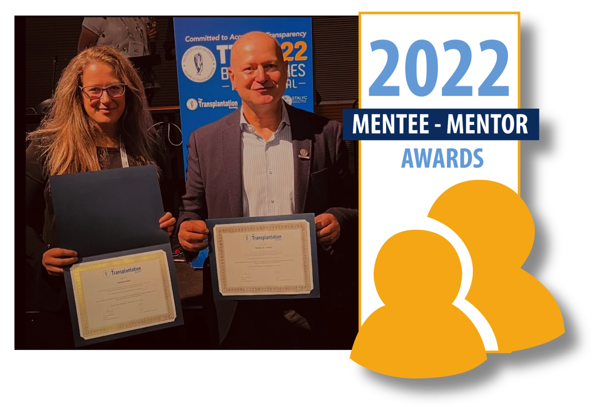
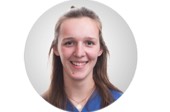
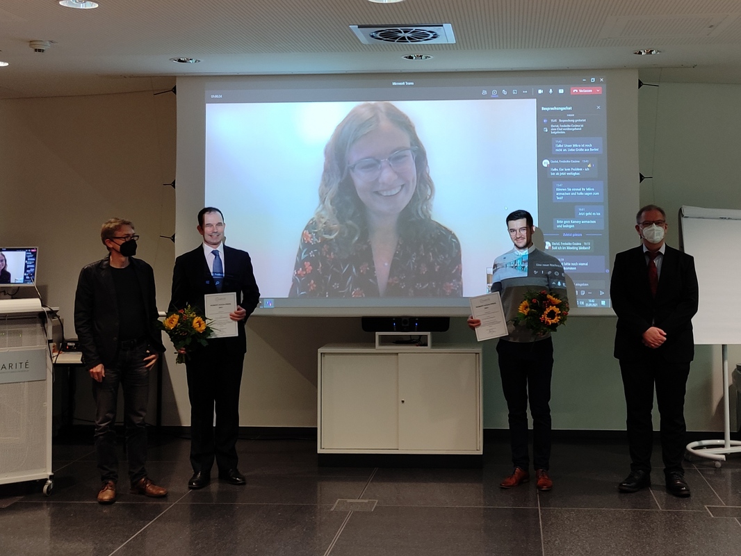
Dr. Barbara Kern und Prof. Dr. Stefan G. Tullius received the 2022 Mentee-Mentor-Award of The Transplant Society during the 29th Conference in Buenos Aires.
In collaboration with National and International Societies, TTS acknowledges and recognizes the efforts of scientists who have advanced our understanding of transplantation science and fostered the development of young investigators.
The Mentee-Mentor Awards are designed to encourage dialogue and interactions between trainees and established investigators, and provide financial support for their joint participation in the Congress.
Congratulations!