Implantation of a Neo Bile Duct in domestic pigs
Stacks Image 29474
European Surgical Research accepted our latest paper entitled "Implantation of a tissue engineered Neo Bile Duct in domestic pigs" for publication. Authors are B. Struecker, K. Hillebrandt, N. Raschzok, K. Jöhrens, A. Butter, P. Tang, A. Andreou, H. Napierala, D. Polenz, A. Reutzel-Selke, T. Denecke, J. Pratschke, and I.M Sauer.

Extrahepatic bile duct injuries remain severe complications during cholecystectomy and often require reconstruction by bilioenteric anastomosis (i.e. hepatico-jejunostomy), which comes along with further long-term complications (e.g. recurring ascending cholangitis, secondary biliary cirrhosis). Furthermore, in case of inherent extrahepatic biliary atresia or during liver transplant artificial or engineered bile ducts could enable novel surgical strategies without the need for hepatico-jejunostomy. We present data on the implantation of in vitro generated Neo Bile Ducts in five domestic pigs. Neo Bile Ducts were engineered through decellularization of allogeneic blood vessels and recellularization with autologous cholangiocytes.On postoperative days 0, 1, 7 and 14 blood samples were taken and analyzed (AST, ALT, Bilirubin, Alkaline Phosphatase, Creatinine and Leukocytes). An magnetic resonance cholangiography was performed on postoperative day 14 with one pig. 14 days after implantation pigs were sacrificed and bile ducts were explanted. All pigs survived the complete study period without severe complications. None of the pigs showed signs of biliary leakage or peritonitis. Neo Bile Ducts were infiltrated by neutrophils and neo-angiogenesis was observed around and into the implanted tissue. Whether the presented technique enables the long-term replacement of native bile ducts has to be further evaluated.
Read More
Human hepatocyte isolation – new paper
Stacks Image 29484
Tissue Engineering, Part C: Methods accepted our paper „Human hepatocyte isolation: Does portal vein embolization affect the outcome?“ Authors are Martin Kluge, Anja Reutzel-Selke, Hendrik Napierala, Karl H. Hillebrandt, Rebeka D. Major, Benjamin Struecker, Annekatrin Leder, Jeffrey Siefert, Peter Tang, Steffen Lippert, Daniel Seehofer, Johann Pratschke, Igor M. Sauer und Nathanael Raschzok.

Primary human hepatocytes are widely used for basic research, pharmaceutical testing, and therapeutic concepts in regenerative medicine. Human hepatocytes can be isolated from resected liver tissue. Preoperative portal vein embolization (PVE) is increasingly used to decrease the risk of delayed postoperative liver regeneration by induction of selective hypertrophy of the future remnant liver tissue. The aim of this study was to investigate the effect of PVE on the outcome of hepatocyte isolation. Primary human hepatocytes were isolated from liver tissue obtained from partial hepatectomies (n=190) using the two-step collagenase perfusion technique followed by Percoll purification. Of these hepatectomies, 27 isolations (14.2%) were performed using liver tissue obtained from patients undergoing PVE prior to surgery. All isolations were characterized using parameters that had been described in the literature as relevant for the outcome of hepatocyte isolation. The PVE and non-PVE groups were similar in regard to donor parameters (sex, age, indication for surgery), isolation parameters (liver weight, cold ischemic time), and the quality of the liver tissue. The mean initial viable cell yield did not differ between the PVE and non-PVE groups (10.16±2.03x10⁶ cells/g vs. 9.70±0.73 x10⁶ cells/g, p=0.499). The initial viability was slightly better in the PVE-group (77.8 ±2.03% vs. 74.4 ±1.06%). The mean viable cell yield (p=0.819) and the mean viability (p=0.141) after Percoll purification did not differ between the groups. PVE had no effect on enzyme leakage and metabolic activity of cultured hepatocytes.  Although PVE leads to drastic metabolic alterations and changes in hepatic blood flow, embolized liver tissue is a suitable source for the isolation of primary human hepatocytes and is equivalent to untreated liver tissue in regard to cell yield and viability.
Read More
Dr. Jan Schröder
Today, Jan Schröder successfully defended his thesis (summa cum laude) entitled "Vergleichende in vivo und in vitro Analysen im Rahmen der Etablierung der kombinierten Leber- und Leberzelltransplantation im Rattenmodell" !

Congratulations !
Read More
Karl Hillebrandt – YPT Rising Star
Stacks Image 29498

Karl Hillebrandt won this year's YPT Rising Stars Video Session Award. He presented the studies on „Optimized decellularization of rat livers by arterial and portal venous perfusion under oscillating pressure conditions“ during the 17th congress of the European Society for Organ Transplantation (ESOT) 2015 in Brüssel. Young Professionals in Transplantation (YPT) is a forum for junior professionals throughout Europe working in the field of transplantation.

Read More
The Morning After
Referring to our paper „CD44 and CXCL9 serum protein levels predict the risk of clinically significant allograft rejection after liver transplantation“ Geoffrey W. McCaughan, Patrick Bertolino and David G. Bowen wrote an interesting editorial entitled „Could The Morning After liver transplant be immunologically interesting?“ 

They conclude, „that our study urges us to study the immune system response in liver allograft recipients during the very early phases after liver transplantation and to explore how events in immune organs and the allograft are reflected within the serum. Whether the patterns observed truly represent early detection of ACR versus tolerance, or a combination of both, requires further study and experimentation, including the identification of the cellular sources of these and other potential markers of immune outcome. It seems that despite significant levels of immunosuppressive drugs, immune activation and engagement occurs very early after human liver transplant, within the first 24 hours, in a manner that may have similarities with experimental animal models. Thus, the morning after effect could be an exciting window to longer-term immune outcomes, rather than just being preoccupied with observing important routine outcomes and detecting early complications.“
Read More
Publication in Jove
Procedure for Decellularization of Rat Livers in an Oscillating-pressure Perfusion Device“ is available in J Vis Exp. 2015 Aug 10;(102). doi: 10.3791/53029 – accessible via the JOVE servers. 

Authors are K. Hillebrandt, D. Polenz, A. Butter, P. Tang, A. Reutzel-Selke, A. Andreou, H. Napierala, N. Raschzok, J. Pratschke, I.M. Sauer, and B. Struecker . The presented techniques for liver harvesting, cannulation and perfusion using our proprietary device enable sophisticated perfusion set-ups to improve decellularization and recellularization experiments in rat livers.
Read More
microRNAs in liver tissue engineering
Stacks Image 29528
Our paper "microRNAs in liver tissue engineering - New promises for failing organs"was accepted for publication in Advanced Drug Delivery Reviews (IF: 15.038). Authors are Nathanael Raschzok, Hannes Sallmon, Johann Pratschke and Igor M. Sauer.

miRNA-based technologies provide attractive tools for several liver tissue engineering approaches. Herein, we review the current state of miRNA applications in liver tissue engineering. Several miRNAs have been implicated in hepatic disease and proper hepatocyte function. However, the clinical translation of these findings into tissue engineering has just begun. miRNAs have been successfully used to induce proliferation of mature hepatocytes and improve the differentiation of hepatic precursor cells. Nonetheless, miRNA-based approaches beyond cell generation have not yet entered preclinical or clinical investigations. Moreover, miRNA-based concepts for the biliary tree have yet to be developed. Further research on miRNA based modifications, however, holds the promise of enabling significant improvements to liver tissue engineering approaches due to their ability to regulate and fine-tune all biological processes relevant to hepatic tissue engineering, such as proliferation, differentiation, growth, and cell function.
Read More
CD44 and CXCL9 predicting rejection after LTx
Stacks Image 29538
Based on a fruitful collaboration with the department of Visceral, Transplantation, Thoracic, and Vascular Surgery at the University of Leipzig our paper on „CD44 and CXCL9 serum protein levels predict the risk of clinically significant allograft rejection after liver transplantation“ has been accepted for publication in Liver Transplantation. 

Authors are Nathanael Raschzok, Anja Reutzel-Selke, Rosa Bianca Schmuck, Mehmet Haluk Morgul, Ulrich Gauger, Kukuh Aji Prabowo, Laura-Marie Tannus, Annekatrin Leder, Benjamin Struecker, Sabine Boas-Knoop, Michael Bartels, Sven Jonas, Christian Lojewski, Gero Puhl, Daniel Seehofer, Marcus Bahra, Andreas Pascher, Johann Pratschke, and Igor Maximilian Sauer.

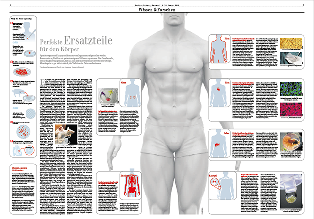
The diagnosis of acute cellular rejection (ACR) after liver transplantation is based on histological analysis of biopsies because non-invasive biomarkers for allograft rejection are not yet established for clinical routines. CD31, CD44, and CXCL9 have previously been described as biomarkers for cross-organ allograft rejection. Here, we assessed the predictive and diagnostic value of these proteins as serum biomarkers for clinically significant ACR in the first six months after liver transplantation in a prospective study. The protein levels were measured in 94 patients immediately prior to transplantation, at postoperative days (POD) 1, 3, 7, and 14, and when biopsies were performed during episodes of biochemical graft dysfunction. Our results suggest that CD44 and CXCL9 may serve as predictive biomarkers to identify liver allograft recipients at risk for clinically significant ACR.
Read More
Cover – march issue of Tissue Engineering
Stacks Image 29543

One of the figures of our paper „Porcine liver decellularization under oscillating pressure conditions – A technical refinement to improve the homogeneity of the decellularization process“ made it to the cover of the march issue of Tissue Engineering, Part C : Methods.

Congratulations to Dietrich Polenz, who made the corrosion cast of a decellularized pig liver matrix: red, hepatic artery; blue, portal vein; yellow, bile duct and gallbladder.

Read More
Particles for microRNA-targeted manipulation
Biomaterials accepted our paper on „Micron-sized iron oxide-containing particles for microRNA-targeted manipulation and MRI-based tracking of transplanted cells“. Authors are Annekatrin Leder, Nathanael Raschzok, Christian Schmidt, Duygu Arabacioglu, Antje Butter, Susanne Kolano, Luisa S. de Sousa Lisboa, Wiebke Werner, Dietrich Polenz, Anja Reutzel-Selke, Johann Pratschke, and Igor M. Sauer.

Particle-based delivery systems for therapeutic manipulation and tracking of transplanted cells by magnetic resonance imaging (MRI) are commonly based on nanometer-sized superparamagnetic iron oxide particles (SPIOs). Here, we present a proof of concept for multifunctional, silica based micron-sized iron oxide-containing particles (sMPIO) that combine fluorescence imaging, MRI tracking, and on-the-spot targeting of specific microRNAs on a particle surface for therapeutic manipulation by RNA interference. Antisense locked nucle-LNA) were covalently bound to the surface of silica-based, DAPI-integrated, micron-sized iron oxide particles (sMPIO--LNA). In vitro studies using primary human hepatocytes showed rapid particle uptake (4 hours) that was accompanied by significant depletion of the targeted microRNA Let7g (80%), up- regulation of the target proteins Cyclin D1 and c-Myc, and specific proteome changes. sMPIO--LNA- labeled cells were successfully detected by fluorescence imaging and could be visualized by MRI after intrasplenic transplantation in rats. This new theranostic particle provides a promising tool for cell transplantation where cellular imaging and microRNA-based manipulation is needed.

Biomaterials is the leading journal in its field. Impact factors released by ISI in July 2014 showed Biomaterials with an impact factor of 8.312!
Read More
Decellularization of pancreata – EPITA Award
Stacks Image 29564
Ben Strücker presented our latest results on DECELLULARIZATION OF WHOLE RAT PANCREATA – EVALUATION OF THREE DIFFERENT PERFUSION ROUTES at the 5th EPITA Winter Symposium in Innsbruck from the 25th to the 27th of January 2015. He received the AIDPIT&EPITA Award for the best oral presentation. Congratulations!

Ben Strücker presented three effective protocols for rat pancreas perfusion decellularization, evaluating different perfusion routes. In contrast to liver decellularization the perfusion route seems to have no major impact on decellularization results. The dPECMs could serve for cellular repopulation with islets from a different (xenogene) origin to generate functional, transplantable endocrine pancreata in vitro.
Read More
Rebeka Major: BIH Promotionsstipendium
Rebeka Major successfully applied for the BIH-Promotionsstipendium grant. Her work will focus in the role of the INDY („I’m Not Dead Yet“) gene in liver regeneration – a project in cooperation with Prof. A. Birkenfeld, Universitätsklinikum Carl Gustav Carus in Dresden.
In the Berlin Institute of Health (BIH), the Max Delbrück Center for Molecular Medicine (MDC) and the Charité - Universitätsmedizin Berlin have joined forces. The core idea is to combine translational research with an overarching systems medicine approach to bridge the gap between basic research and clinical application.
Read More
Presentations at DTG 2014
Stacks Image 29588
Nathanael Raschzok presented his poster „Protein biomarkers for diagnosis and prediction of acute cellular rejection after liver transplantation“, Benjamin Strücker his work entitled „Oszillierende Druckschwankung verbessern signifikant die Ratten- und Schweineleber Dezellularisierung“ and Rosa Schmuck her posters „Risk of postoperative infections after LTX rises with MELD score“ and „Protein biomarkers in bile as a diagnostic tool after liver transplantation“.
Read More
Ben Strücker: Charité Clinical Scientist
Stacks Image 29598
Dr. med Benjamin Strücker successfully applied for the Charité Clinical Scientist 2015 program.
His project is entitled „Humanized Porcine Liver““. Clinical mentor is Prof. Dr. Johann Pratschke, scientific mentors is Priv.-Doz. Dr. med Igor M. Sauer.

The program is supported by Stiftung Charité which was endowed by the entrepreneur Johanna Quandt in order to promote biomedical "knowledge entrepreneurs" that is, change makers in biomedicine at the Charité. The goal of this program is to develop new career paths in clinical specialist medical training. The focus of the training program "Clinical Scientist" is translational research ("bench-to-bedside") which will be realized by a reduction in clinical routine and an improved curriculum with defined goals.
Read More
Sham Laparotomy and microRNA Expression in Rats
Stacks Image 29608
BMC Research Notes accepted our latest paper on „Independent Effects of Sham Laparotomy and Anesthesia on Hepatic microRNA Expression in Rats“ for publication.

Studies on liver regeneration following partial hepatectomy (PH) have identified several microRNAs (miRNAs) that show a regulated expression pattern. These studies involve major surgery to access the liver, which is known to have intrinsic effects on hepatic gene expression and may also affect miRNA screening results. We performed two-third PH or sham laparotomy (SL) in Wistar rats to investigate the effect of both procedures on miRNA expression in liver tissue and corresponding plasma samples by microarray and qRT-PCR analyses. As control groups, non-treated rats and rats undergoing anesthesia only were used. We found that 49 out of 323 miRNAs (15%) were significantly deregulated after PH in liver tissue 12 to 48 hours postoperatively (>20% change), while 45 miRNAs (14%) were deregulated following SL. Out of these miRNAs, 10 miRNAs were similarly deregulated after PH and SL, while one miRNA showed opposite regulation. In plasma, miRNA upregulation was observed for miR-133a and miR-133b following PH and SL, whereas miR-100 and miR-466c were similarly down-regulated following anesthesia and surgery. We show that miRNAs are indeed regulated by sham laparotomy and anesthesia in rats. These findings illustrate the critical need for finding appropriate control groups in experimental surgery.

Authors are W. Werner, H. Sallmon, A. Leder, S. Lippert, A. Reutzel-Selke, M.H. Morgül, S. Jonas, S. Dame, P. Neuhaus, J. Iacomini, S.G. Tullius, I.M. Sauer and N. Raschzok.
Read More
Textbook of Organ Transplantation Set
Stacks Image 29618
Brought to you by the world’s leading transplant clinicians, Textbook of Organ Transplantation provides a complete and comprehensive overview of modern transplantation in all its complexity, from basic science to gold-standard surgical techniques to post-operative care, and from likely outcomes to considerations for transplant program administration, bioethics and health policy.
Beautifully produced in full color throughout, and with over 600 high-quality illustrations, it successfully
- Provides a solid overview of what transplant clinicians/surgeons do, and with topics presented in an order that a clinician will encounter them.
- Presents a holistic look at transplantation, foregrounding the interrelationships between transplant team members and non-surgical clinicians in the subspecialties relevant to pre- and post-operative patient care, such as gastroenterology, nephrology, and cardiology.
- Offers a focused look at pediatric transplantation, and identifies the ways in which it significantly differs from transplantation in adults.
- Includes coverage of essential non-clinical topics such as transplant program management and administration; research design and data collection; transplant policy and bioethical issues.

Editors are Allan D. Kirk, Stuart J. Knechtle, Christian P. Larsen, Joren C. Madsen, Thomas C. Pearson, and Steven A. Webber.
I.M. Sauer, N. Raschzok und P. Neuhaus contributed chapter 47: „Artificial Liver, In Vivo Tissue Engineering, and Organ Printing – Solutions for Organ Scarcity

The Wiley-Blackwell book (ISBN: 978-1-118-87014-3, 1880 pages) is available here.
Read More
Decellularization of porcine livers
Stacks Image 29626
Ben Strücker’s paper entitled „Porcine liver decellularization under oscillating pressure conditions – A technical refinement to improve the homogeneity of the decellularization process“ has been accepted for publication in Tissue Engineering, Part C: Methods.
Co-authors are K. Hillebrandt, R. Voitl, A. Butter, R.B. Schmuck, A. Reutzel-Selke, D. Geisel, K. Joehrens, P.A. Pickerodt, N. Raschzok, G. Puhl, P. Neuhaus, J. Pratschke, and I.M. Sauer.

Decellularization and recellularization of parenchymal organs may facilitate the generation of autologous functional liver organoids by repopulation of decellularized porcine liver matrices with induced liver cells. We present an accelerated (7 h overall perfusion time) and effective protocol for human scale liver decellularization by pressure-controlled perfusion with 1% Triton X-100 and 1% SDS via the hepatic artery (120 mmHg) and portal vein (60 mmHg). In addition, we analyzed the effect of oscillating pressure conditions on pig liver decellularization (n=12). The proprietary perfusion device used to generate these pressure conditions mimics intra-abdominal conditions during respiration to optimize microperfusion within livers and thus optimize the homogeneity of the decellularization process. The efficiency of perfusion decellularization was analyzed by macroscopic observation, histological staining (H&E, Sirius red, Alcian blue), immunohistochemical staining (collagen IV, laminin, fibronectin) and biochemical assessment (DNA, collagen, glycosaminoglycans) of decellularized liver matrices. The integrity of the extracellular matrix post-decellularization was visualized by corrosion casting and three-dimensional CT scanning. We found that livers perfused under oscillating pressure conditions (P+) showed a more homogenous course of decellularization and contained less DNA compared to livers perfused without oscillating pressure conditions (P-). Microscopically, livers from the (P-) group showed remnant cell clusters, while no cells were found in livers from the (P+) group. The grade of disruption of the ECM was higher in livers from the (P-) group, although the perfusion rates and pressure did not significantly differ. Immunohistochemical staining revealed that important matrix components were still present after decellularization. Corrosion casting showed an intact vascular (portal vein and hepatic artery) and biliary framework. In summary, the presented protocol for pig liver decellularization is quick (7 h) and effective. The application of oscillating pressure conditions improves the homogeneity of perfusion and thus the outcome of the decellularization process.
Read More
25th European Student´s Conference
Stacks Image 29641
The ESC is one of the largest student-run biomedical conferences worldwide. It was established in 1989 as one of the first student conferences facilitating exchange between Western and Eastern Europe. Until today, it aims to foster international understanding within the young scientific community, and to promote high-quality science among young researchers in the biomedical field.

This year’s lecture series “Rethinking Medical Research – how do we achieve innovation?” will explore the dichotomy of medical advances by looking at both the opportunities and challenges that are presented to researchers and physicians. With topics ranging from open access policies, pharmaceutical innovations, and revolutionary health policies to advances in global health – we hope that this year’s topic will both challenge and inspire our participants!
The conference will take place from September 17th – 20th at the Virchow Klinikum Campus of the Charité Universitätsmedizin in Berlin, Germany.

More information here!
Read More
N. Raschzok Guest Editor - Call for Papers
Stacks Image 29651
Pluripotent stem cells have been an important tool for researches in hepatology, starting with mouse embryonic stem cells and transgenesis. Together, with other animal models, they largely contributed to our current knowledge in hepatic cells development and disease modeling. Therefore, with the possibility to manipulate human embryonic stem cells and more recently human induced pluripotent stem cells, there was an existing substratum to study these processes in a human environment, which contributed to the tremendous explosion of the emerging Liver Regenerative Medicine field. The use of such cells in the last few years has been already at the origin of numerous breakthroughs in disease modeling, host-pathogen interactions studies, or liver bioengineering and many are to come.

Investigators are invited to participate in a special issue on „Human Pluripotent Stem Cells in Hepatic Development, Liver Reconstruction and Disease Modeling“ through original research articles at the forefront of this fast pace moving field as well as reviews describing current and forthcoming challenges.

Lead Guest Editor is Karim Si-Tayeb (Institute of the Thorax, Nantes). Guest Editors are Gareth Sullivan (Institute of Basic Medical Sciences, University of Oslo), Robert Schwartz (Weill Cornell Medical College, Cornell University, New York), Nathanael Raschzok (Charité – Universitaetsmedizin Berlin), and Tamir Rashid (King's College London).

Manuscript Due: Friday, 23 January 2015
First Round of Reviews: Friday, 17 April 2015
Publication Date: Friday, 12 June 2015
Read More
Kristina Kähm - Bachelor of Science
Kristina Kähm successfully completed her bachelor thesis entitled “Analyse von Glykosaminoglykanen und Fibronektinen in der extrazellulären Matrix zum Nachweis der erfolgten Dezellularisierung von Rattenleber-Explantaten“ and is now a Bachelor of science.
Her project was supervised in cooperation with Prof. Dr. Marcus Frohme, Division Molecular Biotechnology and Functional Genomics, Technical University of Applied Sciences in Wildau, Germany.
Read More
Dr. med. Carolin M. Langer
Stacks Image 29670
Today, Carolin Langer successfully defended her thesis entitled Evaluierung eines Silizium-basierten Eisenoxidpartikels als intrazelluläres Magnetresonanzkontrastmittel für die Leberzelltransplantation „magna cum laude“.

Cellular therapies require methods for noninvasive visualization of transplanted cells. Micron-sized iron oxide particles (MPIOs) generate a strong contrast in magnetic resonance imaging (MRI) and are therefore ideally suited as an intracellular contrast agent to image cells under clinical conditions. However, MPIOs were previously not applicable for clinical use. her thesis focussed on the development and evaluation of silica-based micron-sized iron oxide particles (sMPIOs) with a functionalizable particle surface. Labeling was stable and had no adverse effects on labeled cells. Silica is a biocompatible material that has been approved for clinical use. sMPIOs could therefore be suitable for future clinical applications in cellular MRI, especially in settings that require strong cellular contrast. Moreover, the particle surface provides the opportunity to create multifunctional particles for targeted delivery and diagnostics.

Congratulations !
Read More
Decellularization and oscillating pressure conditions
Stacks Image 29680
The Journal of Tissue Engineering and Regenerative Medicine accepted our latest paper on „Improved rat liver decellularization by arterial perfusion under oscillating pressure conditions“ for publication. Authors are B. Struecker, A. Butter, K. Hillebrandt, D. Polenz , A. Reutzel-Selke, P. Tang, S. Lippert, A. Leder, S. Rohn, D. Geisel, T. Denecke, K. Aliyev, K. Jöhrens, N. Raschzok, P. Neuhaus, J. Pratschke and I.M. Sauer.

One approach of regenerative medicine to generate functional hepatic tissue in vitro is de- and recellularization and several protocols for the decellularization of different species have been published. To the best of our knowledge this is the first report on rat liver decellularization by perfusion via the hepatic artery under oscillating pressure conditions, intending to optimize microperfusion and minimize damage to the ECM. Four decellularization protocols were compared: perfusion via the portal vein (PV) or the hepatic artery (HA), with (+P) or without (-P) oscillating pressure conditions. All rat livers (n=24) were perfused with 1% Triton X-100 and 1% SDS, each for 90 minutes with a perfusion rate of 5ml/min. Perfusion decellularization was observed macroscopically and the decellularized liver matrices (DLMs) were analyzed by histology and biochemical analyses (e.g., levels of DNA, glycosaminoglycans (GAG), and hepatocyte growth factor (HGF). Livers decellularized via the hepatic artery and under oscillating pressure showed a more homogenous decellularization and less remaining DNA, compared to livers of the other experimental groups. The novel decellularization method described is effective, quick (3 hours) and gentle to the ECM and thus represents an improvement of existing methodology.
Read More
<<  Page 9 / 12  >>
Year
© 2025 Prof. Dr. Igor M. Sauer | Charité - Universitätsmedizin Berlin | Disclaimer

This website or its third-party tools use cookies, which are necessary to its functioning and required to achieve the purpose illustrated in the Disclaimer. By closing this banner, scrolling this page, clicking a link or continuing to browse otherwise, you agree to the use of cookies.